Papillary muscles of the heart function. Leaf valves: structure and characteristics. Other questions from the category
The valve apparatus, which is in anatomical and functional unity, consists of correlative connections with other components of the heart, as a result of which, as a result of these correlations that arise both in the embryonic and postnatal periods, significant changes in its design occur with age, which deepen the existing individual , typical and age differences.
Left atrioventricular valve
The mitral valve apparatus is a complex complex structure, the morphological elements of which are the connective tissue atrioventricular ring, leaflets, papillary muscles and chordae tendineae.
Functionally, the mitral valve apparatus also includes the left atrium and left ventricle. The normal function of the valve depends on both the anatomical and functional usefulness of all its elements.
The mitral valve consists of two main leaflets: a large anterior one (aortic, or septal) and a smaller posterior one (mural). The posterior valve usually consists of three or more lobes (scallops), which in the fetus are still separated by subcommissures.
The valves and lobules develop variably in each individual. The number of valves varies: 2 valves in 62% of people, 3 in 19%, 4 in 11% and 5 in 8% of people.
The line of attachment of the anterior leaflet occupies less than half the circumference of the annulus fibrosus. Most of its circumference is occupied by the posterior valve. The front flap is square or triangular in shape and has a larger area than the rear one. The wide and mobile anterior leaflet plays the main role in the closing function of the mitral valve, and the posterior leaflet plays a predominantly supporting function.
Histologically, the mitral valve leaflets consist of three layers: 1) a fibrous layer consisting of dense collagen, continuous with the chordae tendineae; 2) the spongy layer, which is located on the side of the atrial surface and forms the anterior edges of the leaflet (it consists of a small number of collagen fibers and an abundance of proteoglycans, elastin and connective tissue cells); 3) fibroelastic layer covering most valves. The fibroelastic layer thickens with age due to increased production of elastin and collagen; similar changes are also observed in myxomatous mitral valve degeneration.
The epicardial fibers in the left ventricle, emanating from the base of the heart, descend to the apex and are introduced into the cavity in the form of two papillary muscles, which have a vertical orientation of the myocardial fibers. The anterolateral papillary muscle usually has one large head (papilla) and a more developed muscle structure. The posteromedial papillary muscle may have two or more nipples. The structure of papillary muscles is varied. Muscles can have a common base and several apices or one apex and a divided base.
The average distance from the papillary muscles to the mitral annulus is 23.5 mm. The posteromedial papillary muscle is usually supplied by the right coronary artery (in 10% of cases by the left circumflex artery). The anterolateral papillary muscle is supplied by the left descending and circumflex coronary arteries.
During diastole, papillary muscles are visible in the left ventricular inflow tract. During systole they are detected in the output tract. By contracting, the papillary muscles increase left ventricular output. In diastole, papillary muscles make up 5-8% of the volume of the left ventricle, while in systole - 15-30%. The anterior and posterior papillary muscles contract simultaneously and are innervated by both sympathetic and parasympathetic nerves.
A dense network of chordae tendineae extends from the papillary muscles to both mitral leaflets. Chordae are classified into three functional groups. The first group (primary) are chords located in close proximity to the papillary muscles. They progressively separate and attach to the main edges of the valves. Primary chordae are fundamental in preventing valve prolapse in systole. The second group (chords of the second order) are supporting. These chords branch and attach to the ventricular surface of the leaflets in the area of the transition of the tuberous zone to the smooth zone and, thus, form the edges corresponding to the border of cooptation of the leaflets. The second-order chordae play an important role in optimizing left ventricular systolic function. The third group (tertiary or basal) arises from the trabeculae of the left ventricle and has a fan-shaped shape. Additionally, a distinction is made between commissural chords and split chords.
The chordae contain nerve fibers and some (“immature”) chordae may contain muscle fibers. The chordal apparatus consists of approximately 25 main (from 15 to 32) chordal branches arising from the papillary muscles, which, dividing at the valves, form over 100 small chords. Chordae have a differentiated microstructure depending on the type. The presence of vessels in the chords characterizes them as an integral component that coordinates the work of the subvalvular apparatus. The main chords of the anterior mitral leaflet are more vascularized than the other chords. The anterior and posterior marginal chordae contain higher amounts of deoxyribonucleic acid and collagen compared to other chordae.
The mitral valve is a developing structure. Changes in structure and function occur according to the needs of the circulatory system. The increasing load on the body with age causes anatomical and functional restructuring of the valve, aimed primarily at improving its obturator function.
Tricuspid valve
In children under 1 year of age, the diameter of the right atrioventricular orifice is 0.8 - 1.7 cm (usually 1.2-1.5), up to 6 years - 1.7-2.6 cm (usually 2.0-2. 3), up to 12 years - 2.3-3.1 cm (usually 2.5-2.8), up to 17 years - 2.6-3.6 cm (usually 2.7-3.0). In boys, the diameter of the hole is 0.1-0.5 cm larger than in girls.
The number of leaflets in the right atrioventricular valve in children ranges from 2 to 4. With age, the number of leaflets increases. Obviously, in the postnatal period, valve restructuring also occurs, and the formation of additional leaflets is an adaptive mechanism, the purpose of which is to improve the obturator function of the valve.
Usually there are three main leaflets - anterior, posterior and septal, which are observed in 55.7% of cases. In children, an additional anterior leaflet occurs in 7.5% of cases, a posterior leaflet in 21%, and a septal leaflet in 3% of cases.
The sizes of the valves vary individually. The anterior leaf has the largest dimensions. In children, the width of the anterior valve is 0.7-4.5 cm, height - 0.4-2.7 cm. The width of the septal valve is 0.6-3.0 cm, height 0.4-2.0 cm. Width rear flap - 1.6-4.5 cm, height - 1.4-3.0 cm.
Additional doors are smaller in size compared to the main ones and, as a rule, have a triangular shape. In children, their width is 0.4-2.5 cm, height 0.4-2.2 cm.
A comparison of data on the number of leaflets and their sizes with data on the circumference of the right atrioventricular orifice revealed that with a larger circumference, larger leaflets and a greater number of them are more common. With a small circumference of the right atrioventricular orifice, there are usually 3 leaflets with a small width and height.
The papillary muscles, being a continuation of the muscles of the right ventricle, can have a variety of shapes. In the right ventricle one can distinguish papillary muscles of a cylindrical, conical shape, in the form of a truncated tetrahedral pyramid. Papillary muscles can have several heads (multi-headed). The number of papillary muscles in the right ventricle ranges from 2 to 11. In children, the number of anterior papillary muscles ranges from 1 to 3, posterior papillary muscles - from 1 to 4. The number of septal papillary muscles in children and adults varies from 0 to 5. In children, In 3.5% of cases, the posterior papillary muscles are absent, in 6% - the septal muscles. With age, the number of papillary muscles in the right ventricle decreases, which is associated with the fusion of individual muscles into compact, irregularly shaped muscles with several heads. With age, some muscles lag behind the growth of the heart, shorten and even disappear. The anterior papillary muscles are the largest, and the septal muscles are the smallest. In children, the length of the anterior papillary muscles is 0.6-2 cm, the posterior ones - 0.3-1.4 cm, the septal muscles - 0.2-0.8 cm. The length of the papillary muscles of the right ventricle is related to the length of the heart: long papillary muscles are observed on long hearts, short ones on short ones.
The chordae tendineae begin from the papillary muscles, which are attached to the valves along their free edge, as well as along the entire ventricular surface up to the fibrous ring. The number of tendon chords extending from the anterior papillary muscles in children ranges from 5 to 16. 4 to 16 chords extend from the posterior papillary muscles, and from 1 to 13 chords from the septal papillary muscles. There were from 3 to 15 parietal chordae in children.
Analysis of the data obtained on the structure of the tricuspid valve allowed S.S. Mikhailov to identify two extreme forms of its structure. A simple form of the tricuspid valve structure is observed with a narrow and long heart in each age group. With this form of valve structure, the diameter of the fibrous ring is the smallest (in children under 1 year of age - 0.8-1.2 cm, up to 6 years - 1.7-2.0 cm, up to 12 years - 2.3-2, 8 cm, up to 18 years - 2.6-3.0 cm, in adults - 2.7-3.0 cm), its branches are thin, more often there are 2-3 valves and 2-4 papillary muscles, from which the valves 16-25 chords.
The second form of the tricuspid valve structure is complex. This form is noted on preparations of a wide and short heart. With this form of valve structure, the diameter of the fibrous ring is the largest (in children under 1 year of age - 1.3-1.7 cm, up to 6 years - 2.1-2.6 cm, up to 12 years - 2.9-3, 1 cm, up to 18 years - 3.1-2.6 cm, in adults - 3.6-4.8 cm), its branches are thick, 4-6 valves, 6-10 papillary muscles, 30-40 outgoing chordae.
Aortic valve
The aortic valve is located at the mouth of the aorta and consists of three semilunar cusps attached to the annulus fibrosus. The condition of the latter and the structure of the initial part of the aorta have a direct impact on the function of the valves, therefore the fibrous ring of the aorta and the sinuses of Valsalva are usually classified as components of the aortic valve.
Each leaflet has the appearance of a thin plate, the mechanical basis of which is the fibrous layer, which is a continuation of the fibrous ring of the aorta. On the side of the aorta and ventricle, the fibrous plate is covered by endothelial, subendothelial layers and a layer of elastic fibers.
There are right, left and posterior (non-coronary) cusps of the aortic valve. The places where the valves connect to each other are called commissures. There are anterior commissure (between the right and left valves), right commissure (between the right and posterior valves) and posterior commissure (between the left and posterior valves).
The sizes of the semilunar valves have both age and individual differences. Typically, the width of the semilunar valves exceeds the width of the aortic sinuses, and their height, on the contrary, is less than the height of the aortic sinuses. The width of the semilunar valves in children is: right - from 8.4?2.16 to 17.0?3.1 mm, left - from 7.2?2.2 to 16.0?3.2 mm, back - from 9.00?2.56 to 21.5?1.62 mm; in adults, the right flap is from 25.00?3.53 to 28.0?2.6 mm, the left one is from 22.5?3.1 to 26.0?2.6 mm, the back one is from 26?3 to 28.0?3.2 mm.
The space between the wall of the aortic sinuses and the outer surface of the semilunar valves (facing the sinus wall) is called the sockets of the aortic valves (lunalae valvularum semilunarium). Due to the fact that the semilunar valves are wider than the aortic sinuses, and the height of the valves is less than the height of the sinuses, blood under pressure, when entering the aortic bulb, spreads into the holes of the semilunar valves, displacing them downward, closing the aortic valve.
The aortic crescents are supplied with blood not only due to the oxygenated blood flowing in the aorta, but also due to their own microvascular bed, the condition of which plays an important role in the normal functioning of the valve and in the development of pathological processes.
Pulmonary valve
The pulmonary valve consists of a fibrous ring, the wall of the trunk and three semilunar valves attached to it. In the initial part of the pulmonary trunk, just like in the aorta, there is an expansion in which there are depressions - the sinuses of the pulmonary trunk.
The fibrous ring is located in the same way as in the aorta, on the inner surface of the connection between the wall of the conus arteriosus and the wall of the pulmonary trunk. The semilunar valves of the pulmonary valve originate from the medial edge of the fibrous ring. Fibrous rings covered with endocardium form the bottom of the sinuses of the pulmonary trunk.
The semilunar valves begin from the fibrous ring of the pulmonary trunk and are represented by a fold of the endocardium. There are anterior, left and right semilunar valves of the pulmonary trunk. The lower edges of the valves are fused with the lower edges of the sinuses. There are nodules (noduli) on the upper edges of the flaps. The valves together with the sinuses form the lunula (lunuli). The dimensions of the semilunar valves are slightly larger than the sinuses of the pulmonary trunk.
In parallel with the growth of the heart, the size of the great vessels increases, but their growth rate is slower. So, if the volume of the heart increases 7 times by the age of 15, then the circumference of the aorta increases only 3 times. Over the years, the difference in the size of the lumen of the openings of the pulmonary trunk and the aorta decreases somewhat. If at the time of birth the ratio of the lumens of the pulmonary trunk and aorta exceeds 20-25% (aorta - 16 mm, pulmonary trunk - 21 mm), then by 10-12 years their lumen is comparable, and in adults the lumen of the aorta exceeds the lumen of the pulmonary trunk (aorta - 80 mm, pulmonary trunk - 74 mm). The circumference of the pulmonary trunk in children is always greater than the circumference of the ascending aorta. The lumen of the arteries in general narrows somewhat with age relative to the size of the heart and increasing body length. Only after 16 years does some expansion of the arterial vascular bed occur.
The length of the aorta before the bifurcation at the time of birth is on average 125 mm, its diameter at the exit is about 6 mm. The same width is characteristic of the descending section. The aortic isthmus, located 10 mm from the origin of the left subclavian artery, has an internal diameter of only about 4 mm. In the first months of life, the area of the isthmus expands, and after six months the narrowing of the lumen is no longer detectable here.
The pulmonary trunk is relatively short at the time of birth and is divided into two approximately equal pulmonary arteries, which creates a pressure difference between the vessels in some children, reaching up to 8-15 mm Hg, and can cause the characteristic systolic murmur of peripheral pulmonary artery stenosis. After birth, the lumen of the pulmonary trunk does not initially increase, and the diameter of the pulmonary arteries grows quite intensively, which leads to the disappearance of the pressure drop, usually after 5-6 months. The wall of the pulmonary trunk consists of a framework of elastic fibers alternating with smooth muscle elements. In response to hypoxia and acidosis, the lumen of the artery can significantly decrease. In a child's first weeks and months, the muscular layer of the pulmonary vessels is less pronounced, which explains the smaller response of children to hypoxia.
An important role in functions of atrioventricular valves The apparatus that holds the valves plays - tendon threads, attached, on the one hand, to the free edge of the valve leaflets, and on the other, to the tops of the papillary muscles. With endocarditis and myocarditis, these formations are involved in the process, undergo pathological changes, and therefore, to a greater or lesser extent, disrupt the function of the affected valves to a certain extent.
Tendon threads like valves, they consist of fibrous, cell-poor tissue covered with a very thin layer of endocardium. Passing into the tissue of the valve leaflets, the fibrous tissue of the threads is fan-shaped distributed in the fibrous plate of the leaflet. From each papillary muscle one or more tendon filaments arise, which are attached to the free edges of the valves or, less commonly, to their ventricular surface.
Bunch of threads from each papillary muscle is divided in the left ventricle into two parts, one of which goes to the posterior leaflet, the other to the anterior one. The distribution of attachment of filaments to the leaflets in the right ventricle is not so clearly defined. A large number of thin tendon filaments start directly from the muscles of the ventricles and go to different parts of the valves. The tendon threads of the left ventricle are thicker and more numerous than the right.
If all the threads are the same tense, then the valve flaps are evenly stretched (A. M. Eliseeva, 1948).
Naturally, when endocarditis the inflammatory process can also spread to tendon threads; valve leaflets undergo changes with their tendon threads. As a result of the organization of thrombotic masses and sclerosis, thickening, shortening, coarsening and compaction of tendon threads occurs. Sometimes adjacent tendon threads become fused to each other either due to the organization of thrombotic masses enveloping them, or due to the proliferation of connective tissue on the side of the valves.
In these cases they may form connecting plates like the swimming membranes of a frog's foot. The process of sclerosis can also affect the apices of the papillary muscles. With ulcerative endocarditis, the process moves to the tendon threads, leading to their destruction and rupture. Sometimes all the tendon threads of one leaf are torn.
Important role in ensuring normal The functions of the atrioventricular valves belong to the papillary (papillary) muscles. These muscles are relatively large in the left ventricle, but not in the right. When the papillary muscles are tense, the adjacent edges of the valves move closer together.
For inflammation myocardium the same processes are observed in the papillary muscles. The bases of the papillary muscles are first and most severely affected. Sclerosis and death of muscle tissue are also more pronounced in the papillary muscles than in the rest of the myocardium. This is confirmed by the data of M.A. Skvortsov (1950).
Spreading ulcerative endocarditis or purulent myocarditis can lead to destruction of the papillary muscles and their separation. The same pathology can be caused by an infarction of the ventricular wall with involvement of the papillary muscle or with limited necrosis. Most often this happens to the posterior papillary muscle of the left ventricle. At the autopsy they find a piece of muscle hanging freely on tendon threads (E.M. Gelshtein, 1951).
Break or even full separation of the valve, tendon threads, as well as papillary muscles can be observed without destructive processes in the endocardium or myocardium - due to significant physical stress, sharp bruises and compression of the chest, a fall from a height, or direct injuries to the heart. The same reasons can cause rupture of the walls of the heart and large vessels. Traumatic rupture of the heart occurs especially easily with pathological changes in the myocardium due to myocarditis, cardiosclerosis, aneurysms (V.N. Sirotinin, 1913; A. Fokht, 1920).
Pathological processes, leading to sclerosis (of the valves, orifices, tendon threads and their muscles), underlie the pathology that is commonly called heart defects or, more precisely, heart valve defects. Damage to the valves leads to greater or lesser disruption of the heart, resulting in more or less severe circulatory failure.
The heart has a complex structure and performs an equally complex and important job. Contracting rhythmically, it ensures blood flow through the vessels.
The heart is located behind the sternum, in the middle section of the chest cavity and is almost completely surrounded by the lungs. It may move slightly to the side as it hangs freely on the blood vessels. The heart is located asymmetrically. Its long axis is inclined and forms an angle of 40° with the axis of the body. It is directed from top to right, forward, down to the left, and the heart is rotated so that its right section is tilted more forward, and the left one – back. Two-thirds of the heart is to the left of the midline and one-third (the vena cava and right atrium) is to the right. Its base is turned towards the spine, and its apex is facing the left ribs, to be more precise, the fifth intercostal space.

Sternocostal surface the hearts are more convex. It is located behind the sternum and cartilages of the III-VI ribs and is directed forward, upward, and to the left. A transverse coronary groove runs along it, which separates the ventricles from the atria and thereby divides the heart into an upper part, formed by the atria, and a lower part, consisting of the ventricles. Another groove of the sternocostal surface - the anterior longitudinal - runs along the border between the right and left ventricles, with the right one forming the largest part of the anterior surface, the left one the smaller one.
Diaphragmatic surface flatter and adjacent to the tendon center of the diaphragm. A longitudinal posterior groove runs along this surface, separating the surface of the left ventricle from the surface of the right. In this case, the left one makes up the majority of the surface, and the right one makes up the smaller part.
Anterior and posterior longitudinal grooves they merge at their lower ends and form a cardiac notch to the right of the cardiac apex.
There are also side surfaces located on the right and left and facing the lungs, which is why they are called pulmonary.
Right and left edges hearts are not the same. The right edge is more pointed, the left is more blunt and rounded due to the thicker wall of the left ventricle.
The boundaries between the four chambers of the heart are not always clearly defined. The landmarks are the grooves in which the blood vessels of the heart are located, covered with fatty tissue and the outer layer of the heart - the epicardium. The direction of these grooves depends on how the heart is located (obliquely, vertically, transversely), which is determined by the body type and the height of the diaphragm. In mesomorphs (normosthenics), whose proportions are close to the average, it is located obliquely, in dolichomorphs (asthenics) with a thin physique - vertically, in brachymorphs (hypersthenics) with wide short forms - transversely.

The heart seems to be suspended by the base on large vessels, while the base remains motionless, and the apex is in a free state and can move.
Structure of heart tissue
The heart wall is made up of three layers:
- The endocardium is the inner layer of epithelial tissue that lines the cavities of the heart chambers from the inside, exactly repeating their relief.
- The myocardium is a thick layer formed by muscle tissue (striated). The cardiac myocytes of which it consists are connected by many bridges that link them into muscle complexes. This muscle layer ensures the rhythmic contraction of the chambers of the heart. The myocardium is thinnest at the atria, the greatest is at the left ventricle (about 3 times thicker than the right), since it needs more force to push blood into the systemic circulation, in which the resistance to flow is several times greater than in the small circle. The atrial myocardium consists of two layers, the ventricular myocardium - of three. The atrial myocardium and ventricular myocardium are separated by fibrous rings. The conduction system that provides rhythmic contraction of the myocardium is one for the ventricles and atria.
- The epicardium is the outer layer, which is the visceral lobe of the heart sac (pericardium), which is a serous membrane. It covers not only the heart, but also the initial parts of the pulmonary trunk and aorta, as well as the final parts of the pulmonary and vena cava.

Anatomy of the atria and ventricles
The cardiac cavity is divided by a septum into two parts - right and left, which do not communicate with each other. Each of these parts consists of two chambers - the ventricle and the atrium. The septum between the atria is called the interatrial septum, and the septum between the ventricles is called the interventricular septum. Thus, the heart consists of four chambers - two atria and two ventricles.
Right atrium
It is shaped like an irregular cube, with an additional cavity in front called the right ear. The atrium has a volume of 100 to 180 cubic meters. cm. It has five walls, 2 to 3 mm thick: anterior, posterior, superior, lateral, medial.
The superior vena cava (from above, behind) and the inferior vena cava (from below) flow into the right atrium. On the lower right is the coronary sinus, where the blood of all the cardiac veins drains. Between the openings of the superior and inferior vena cava there is an intervenous tubercle. In the place where the inferior vena cava flows into the right atrium, there is a fold of the inner layer of the heart - the valve of this vein. The sinus of the vena cava is the posterior dilated section of the right atrium, into which both of these veins flow.
The chamber of the right atrium has a smooth internal surface, and only in the right appendage with the adjacent anterior wall the surface is uneven.
Many pinpoint openings of the small veins of the heart open into the right atrium.
Right ventricle
It consists of a cavity and an arterial cone, which is a funnel directed upward. The right ventricle has the shape of a triangular pyramid, the base of which faces upward and the apex faces downward. The right ventricle has three walls: anterior, posterior, medial.
The front is convex, the back is flatter. The medial is the interventricular septum, consisting of two parts. The larger one, the muscular one, is located at the bottom, the smaller one, the membranous one, is at the top. The pyramid faces the atrium with its base and has two openings: posterior and anterior. The first is between the cavity of the right atrium and the ventricle. The second goes into the pulmonary trunk.
Left atrium
It has the appearance of an irregular cube, is located behind and adjacent to the esophagus and the descending aorta. Its volume is 100-130 cubic meters. cm, wall thickness – from 2 to 3 mm. Like the right atrium, it has five walls: anterior, posterior, superior, literal, medial. The left atrium continues anteriorly into an additional cavity called the left appendage, which is directed towards the pulmonary trunk. Four pulmonary veins flow into the atrium (back and above), in the openings of which there are no valves. The medial wall is the interatrial septum. The inner surface of the atrium is smooth, the pectineus muscles are present only in the left appendage, which is longer and narrower than the right one, and is noticeably separated from the ventricle by an interception. It communicates with the left ventricle via the atrioventricular orifice.
Left ventricle
It is shaped like a cone, the base of which faces upward. The walls of this chamber of the heart (anterior, posterior, medial) have the greatest thickness - from 10 to 15 mm. There is no clear boundary between the front and back. At the base of the cone are the openings of the aorta and the left atrioventricular opening.
The round opening of the aorta is located in front. Its valve consists of three valves.
Heart size
The size and weight of the heart varies from person to person. The average values are as follows:
- length is from 12 to 13 cm;
- greatest width – from 9 to 10.5 cm;
- anteroposterior size – from 6 to 7 cm;
- weight in men - about 300 g;
- weight in women is about 220 g.

Functions of the cardiovascular system and heart
The heart and blood vessels make up the cardiovascular system, the main function of which is transport. It consists of supplying tissues and organs with nutrition and oxygen and returning metabolic products.
The heart acts as a pump - it ensures continuous circulation of blood in the circulatory system and delivery of nutrients and oxygen to organs and tissues. Under stress or physical exertion, its work is immediately restructured: it increases the number of contractions.
The work of the heart muscle can be described as follows: its right part (venous heart) receives waste blood saturated with carbon dioxide from the veins and gives it to the lungs to be saturated with oxygen. From the lungs, O2-enriched blood is directed to the left side of the heart (arterial) and from there is forcefully pushed into the bloodstream.
The heart produces two circles of blood circulation - large and small.
The large one supplies blood to all organs and tissues, including the lungs. It begins in the left ventricle and ends in the right atrium.
The pulmonary circulation produces gas exchange in the alveoli of the lungs. It begins in the right ventricle and ends in the left atrium.
Blood flow is regulated by valves: they prevent it from flowing in the opposite direction.
The heart has such properties as excitability, conductivity, contractility and automaticity (excitation without external stimuli under the influence of internal impulses).
Thanks to the conduction system, sequential contraction of the ventricles and atria occurs, and the synchronous inclusion of myocardial cells in the contraction process.
Rhythmic contractions of the heart ensure a portioned flow of blood into the circulatory system, but its movement in the vessels occurs without interruption, which is due to the elasticity of the walls and the resistance to blood flow that occurs in small vessels.
The circulatory system has a complex structure and consists of a network of vessels for different purposes: transport, shunting, exchange, distribution, capacitance. There are veins, arteries, venules, arterioles, capillaries. Together with the lymphatic ones, they maintain the constancy of the internal environment in the body (pressure, body temperature, etc.).
Arteries move blood from the heart to the tissues. As they move away from the center, they become thinner, forming arterioles and capillaries. The arterial bed of the circulatory system transports necessary substances to the organs and maintains constant pressure in the vessels.

The venous bed is more extensive than the arterial bed. Veins move blood from tissues to the heart. Veins are formed from venous capillaries, which merge to first become venules, then veins. They form large trunks near the heart. There are superficial veins, located under the skin, and deep veins, located in the tissues next to the arteries. The main function of the venous section of the circulatory system is the outflow of blood saturated with metabolic products and carbon dioxide.
To assess the functional capabilities of the cardiovascular system and the permissibility of stress, special tests are carried out, which make it possible to assess the performance of the body and its compensatory capabilities. Functional tests of the cardiovascular system are included in the physical examination to determine the degree of fitness and general physical fitness. The assessment is based on such indicators of the heart and blood vessels as blood pressure, pulse pressure, blood flow speed, minute and stroke volumes of blood. Such tests include Letunov's tests, step tests, Martinet's test, Kotov's - Demin's test.
The heart begins to beat from the fourth week after conception and does not stop until the end of life. It does a tremendous job: per year it pumps about three million liters of blood and makes about 35 million heartbeats. At rest, the heart uses only 15% of its resource, and under load – up to 35%. Over an average lifespan, it pumps about 6 million liters of blood. Another interesting fact: the heart supplies blood to 75 trillion cells in the human body, except for the cornea of the eyes.
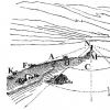
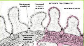
The structure of the leaf valve
Posterior interventricular groove.
The interventricular grooves run from the coronary groove towards the apex of the heart along the anterior and posterior surfaces, respectively, and correspond to the interventricular septum of the heart. The grooves contain the heart's own blood vessels and nerves. This The grooves correspond to septa that divide the heart into 4 sections: longitudinal interatrial and interventricular septa divide the organ into two isolated halves - right and left heart, transverse septum divides each of these halves into the upper chamber - the atrium and the lower chamber - the ventricle of the heart.
The right half of the heart contains venous blood, and the left one – arterial
Structure of the chambers of the heart.
Right atrium is a cavity with a volume of 100-185 ml, resembling a cube in shape, located at the base of the heart on the right and behind the aorta and pulmonary trunk. Separates from the left atrium interatrial septum which shows fossa oval, which is a remnant of an overgrown opening that existed between the two atria during embryogenesis.
The superior and inferior vena cava, coronary sinus and the smallest veins of the heart flow into the right atrium. The upper part of the atrium is atrial appendage , on the inner surface of which longitudinal muscle ridges are visible - the pectineus muscles.
The right atrium communicates with the right ventricle through the right atrioventricular orifice.
Between the latter and the confluence of the inferior vena cava there is the opening of the coronary sinus, and next to it are the pinpoint orifices of the smallest veins of the heart.
Right ventricle . It has the shape of a pyramid with the top facing down. Occupies most of the anterior surface of the heart. It is separated from the left ventricle interventricular septum, most of which is muscular, and the smaller part, located at the very top, closer to the atria, is membranous. At the top two holes in the wall:
1. posterior – right atrioventricular
2. in front – the opening of the pulmonary trunk.
The atrioventricular orifice is closed by the right atrioventricular valve (tricuspid valve),
1. Doors - there are three of them - anterior, posterior, medial, which are triangular tendon plates.
2. Chordae tendineae (threads)
During ventricular systole, the tricuspid valve closes, and the tension of the chordae tendineae prevents the cusps from everting toward the atrium.
Between the ventricle and the pulmonary trunk there is also a valve called semilunar.
The semilunar valve consists of
Front, left and right
Lunar valves,
Arranged in a circle, convex
Surface into the cavity of the right
The ventricle is rather concave and free
The edge is in the lumen of the pulmonary trunk.
When the muscles of the ventricle contract, the semilunar valves are pressed against the wall of the pulmonary trunk by the blood flow and do not interfere with the passage of blood from the ventricle; during relaxation, when the pressure in the ventricular cavity drops, the return flow of blood fills the pockets between the wall of the pulmonary trunk and each of the semilunar valves and opens the valves, their edges close and do not allow blood to pass to the heart.
Left atrium . It has the shape of an irregular cube. Demarcated from the right by the interatrial septum; also has a left ear. In the posterior section of the upper wall, four pulmonary veins open into it, devoid of valves, through which arterial blood flows from the lungs. It communicates with the left ventricle through the left atrioventricular orifice, only near which there are pectineus muscles, the rest of the surface is smooth.
Left ventricle . Cone-shaped, its base faces upward. In the anterior superior section there is the aortic opening, through which the ventricle communicates with the aorta, and the left atrioventricular opening. At the point where the aorta exits the left ventricle there is an aortic valve - the semilunar valve - consisting of right, left, and posterior valves. The aortic valves are thicker than those in the pulmonary trunk.
The left atrioventricular orifice is closed by the leaflet valve, which consists of two leaflets (anterior and posterior) and is therefore also called mitral , chordae tendineae and two papillary muscles.
To diagnose cardiac diseases, a stethoscope is used to auscultation (listening) heart: in this case, the mitral valve is heard at the apex of the heart, the pulmonary trunk and aortic valves are heard in the second intercostal space, respectively, at the right and left edge of the sternum. The place of auscultation (listening) of the tricuspid valve is a point located at the base of the xiphoid process of the sternum.
STRUCTURE OF THE HEART WALL.
The heart wall consists of three layers: the inner - endocardium,
Middle - myocardium, the thickest,
External - epicardium.
1. Endocardium – lines all the cavities of the heart, tightly fused with the underlying muscle layer. On the side of the heart cavities it is lined with endothelium. The endocardium forms the leaflet and semilunar valves.
2. Myocardium is the most powerful and thick wall of the heart. The muscle layer of the walls of the atria is thin due to the low load. Comprises surface layer, common to both atria, and deep- separate for each of them. In the walls of the ventricles it is the most significant in thickness and consists of three layers: the outer - longitudinal, the middle - annular, and the inner longitudinal layers. The muscle layer of the left ventricle is stronger than the right.
The composition of cardiac striated muscle tissue includes typical contractile muscle cells - cardiomyocytes and atypical - cardiac myocytes, which form the conduction system of the heart, ensuring the automaticity of heart contractions, and also coordinates the contractile function of the myocardium of the atria and ventricles of the heart.
3. Epicard – covers the outer surface of the heart and the areas closest to the heart of the aorta and pulmonary trunk, and vena cava. It is part of the fibroserous membrane of the pericardium. In the pericardium there are two layers:
fibrous pericardium, formed by dense fibrous connective tissue, and
serous pericardium, also consisting of fibrous fabric with elastic fibers.
The serous pericardium consists of an internal visceral plate (epicardium), directly covering the heart and tightly connected to it, and an external parietal plate, lining the fibrous pericardium from the inside and passing into the epicardium at the point where large vessels depart from the heart.
The fibrous pericardium at the base of the heart continues into the adventitia of the great vessels; The pleural sacs are adjacent to the pericardium on the side, from below it grows to the tendon center of the diaphragm, and in front it is connected by connective tissue fibers to the sternum.
The pericardium insulates the heart from surrounding organs, and the serous fluid between its plates reduces friction during heart contractions.
Right ventricle of the heart occupies most of the anterior surface of the organ. It has a thicker wall, because there are three layers of myocardium here, and not two, as in the left and right atria. The cavity of this part of the heart has an interesting shape, which could be easily studied if plaster was poured into it and a cast was made. The result would be a kind of “cobblestone” with two spurs. Accordingly, three parts are distinguished in the ventricle (Fig. 1): entrance department(1) - has a short length, but very wide, originates from the atrioventricular opening (2), output department(3), called in older manuals the "arterial sinus" and leading to the pulmonary trunk with its semilunar valve (4), and muscular section(5), occupying the main volume. The inner surface of the muscular section is also smooth due to the endothelium, but not so smooth: from the side of the ventricular wall, fleshy crossbars protrude into the cavity (more often called trabeculae), from the largest of which - the transverse marginal trabecula - the papillary muscles originate. Most often there are three of them: anterior (6), posterior (7) and septal (8), but it happens that there are more of them.
Fig.1. Diagram of the structure of the right ventricle
A very important element structure of the ventricles of the heart are chords - tendon threads(9), or when translated literally from Latin, tendinous strings. These are thin whitish threads originating from the tops of the papillary muscles and ending on the surfaces of the three leaflets of the atrioventricular valves (also, by the way, the anterior, posterior and septal). In this case, there is a kind of overlap. Thus, the anterior papillary muscle “sends” the threads mainly to the anterior of the three valves and partly to the posterior one, the posterior muscle mainly to the posterior valve and partly to the third, septal. Accordingly, from the septal papillary muscle, the tendon threads approach the cusp of the same name of the tricuspid valve and in several bundles - to the anterior one. Output and input departments, divides supraventricular ridge, it flows into the cavity of the left ventricle. The output and input sections are clearly distinguishable, they are smoother on the inside, since the bulk of the trabeculae are in the muscular section. Recall that the right ventricle has two openings: the atrioventricular opening and the pulmonary trunk opening.
The posteroinferior section is represented left ventricle of the heart. Landmarks for the location of the left ventricle can be the diaphragmatic surface, the obtuse edge and the apex of the heart, as well as the left part of the coronary and both interventricular grooves, which are the outer boundaries. Although left ventricle of the heart smaller than the right one, it is not much different from it. There are also three layers of myocardium, however, the wall of the left ventricle is even thicker (1.2 cm) due to a more developed muscle layer. It is worth noting that the wall of the right ventricle has a size of 0.3 cm. In the left ventricle (Fig. 2), the following sections are also distinguished: input(1), that is, closest to the atrioventricular opening (2), day off(3), continuing into the aorta (4), and muscular(5), but in the case of this cardiac cavity there is no such pronounced boundary between the inlet and outlet sections as the supraventricular crest. This is another feature and difference in structure of the ventricles of the heart.

Fig.2. Diagram of the structure of the left ventricle
There is only a fairly conventional delimiter between the inlet and outlet sections, and this is the anterior leaflet (6) of the mitral valve. This delimiter is conditional since it appears as such only during the opening of the valve (Fig. 2, a). If the valve is closed, then there is no anterior leaflet in the cavity, and the division of the ventricle into sections is not noticeable (Fig. 2, b). They go to the mitral valve tendon threads papillary muscles, the most developed are two papillary muscles (or two groups of muscles): anterior (7) and posterior (8), respectively tendon threads These muscles go to the anterior and posterior leaflets of the mitral valve. There are two openings: atrioventricular and aortic. The first is with a bicuspid (mitral) valve. The second is covered by three semilunar valves. The left ventricle sends blood to the aorta through the aortic opening, and then the blood is distributed throughout the body.
Read also...
- Why do you dream about a man’s back?
- Fortune telling with hearts online: a simple and free way to tell fortunes about a guy’s love
- Dream Interpretation: flying above the ground in a dream
- Description of orange zest with photo, its calorie content; how to make at home; use of the product in cooking; harm and beneficial properties