Nephrotic syndrome - the essence and methods of combating the disease. Nephrotic syndrome - causes and symptoms. Symptoms and treatment of acute and chronic nephrotic syndrome Increased blood pressure in nephrotic syndrome is associated
Nephrotic syndrome
What is Nephrotic Syndrome -
Nephrotic syndrome- a nonspecific clinical and laboratory symptom complex, expressed in massive proteinuria (5 g/day or more), disorders of protein-lipid and water-salt metabolism. These disorders are manifested by hypoalbuminemia, dysproteinemia (with a predominance of (Chd-tobulins), hyperlipidemia, lipiduria, as well as edema to the degree of anasarca with dropsy of the serous cavities.
What provokes / Causes of Nephrotic syndrome:
Pathogenesis (what happens?) during Nephrotic syndrome:
Pathogenesis of nephrotic syndrome closely related to the underlying disease. Most of the diseases listed above have an immunological basis, that is, they arise due to the deposition in the organs (and kidney) of complement fractions, immune complexes or antibodies against the glomerular basement membrane antigen with concomitant disorders of cellular immunity.
The main link in the pathogenesis of the leading symptom of nephrotic syndrome - massive proteinuria - is the decrease or disappearance of the constant electrical charge of the wall of the capillary loop of the glomerulus. The latter is associated with the depletion or disappearance of sialoprotein, which normally “dresses” the epithelium and its processes lying on the basement membrane with a thin layer and is part of the membrane itself. As a result of the disappearance of the “electrostatic trap,” proteins are released into the urine in large quantities. Soon there is a “disruption” of the process of protein reabsorption in the proximal tubule of the nephron. Unreabsorbed proteins enter the urine, causing their composition to be selective (albumin and transferrin) or non-selective (high molecular weight proteins, for example, alpha (two)-M G) the nature of proteinuria.
All other numerous disorders in nephrotic syndrome are secondary to massive proteinuria. Thus, as a result of hypoalbuminemia, a decrease in plasma colloid osmotic pressure, hypovolemia, a decrease in renal blood flow, increased production of ADH, renin and aldosterone with hyperresorption of sodium, edema develops.
Histological and cytological studies primarily reveal changes characteristic of nephropathies that caused the development of nephrotic syndrome. The histological signs of nephrotic syndrome itself include fusion of the stalked processes and spreading of podocyte bodies in the glomeruli, hyaline and vacuolar degeneration of proximal tubule cells, and the presence of “foamy” cells containing lipids.
Symptoms of Nephrotic Syndrome:
Clinical picture of nephrotic syndrome, in addition to edema, dystrophic changes in the skin and mucous membranes, can be complicated by peripheral phlobothrombosis, bacterial, viral, fungal infections of various localizations, edema of the brain, retina of the fundus, nephrotic crisis (hypovolemic shock). In some cases, signs of nephrotic syndrome are combined with arterial hypertension (mixed form of nephrotic syndrome).
The course of nephrotic syndrome depends on the form of nephropathy and the nature of the underlying disease. In general, nephrotic syndrome is a potentially reversible condition. Thus, lipoid nephrosis (even in adults) is characterized by spontaneous and drug-induced remissions, although there may be relapses of nephrotic syndrome (up to 5-10 times over 10-20 years). With radical elimination of the antigen (timely surgery for the tumor, exclusion of the antigen drug), complete and stable remission of nephrotic syndrome is possible. The persistent course of nephrotic syndrome occurs in membranous, mesangioproliferative and even fibroplastic glomerulonephritis. The progressive nature of the course of nephrotic syndrome with outcome in chronic renal failure in the first 1.5-3 years of the disease is observed with focal segmental hyalinosis, extracapillary nephritis, subacute lupus nephritis.
Diagnosis of Nephrotic syndrome:
Diagnosis is based on detected changes in blood and urine tests (proteinuria, hyperlipidemia, hypoproteinemia), and on clinical data. The MINS clinic develops gradually, and extrarenal symptoms predominate, especially edematous: increasing swelling appears, first of the eyelids, face, lumbar region (later it can reach the degree of anasarca - widespread swelling of the subcutaneous tissue), genital organs, ascites, hydrothorax, less often - hydropericardium. Characterized by significant hepatomegaly due to liver dystrophy. The skin becomes pale (“pearly” pallor) in the absence of anemia, dry, signs of hypovitaminosis A, C, B1, B2, and degenerative changes appear. Brittleness and dullness of the hair may be observed, and there may be cracks in the skin from which fluid leaks, striae distensae. The child is lethargic, eats poorly, develops shortness of breath, tachycardia, and systolic murmur at the apex (“hypoproteinemic cardiopathy”).
A severe complication in patients with anasarca, i.e., severe hypoproteinemia, can be hypovolemic shock, which is preceded by anorexia, vomiting, and severe abdominal pain. In the observations of N. D. Savenkova and A. V. Papayan (1997), abdominal pain syndrome develops in 23.5% of children with hypoalbuminemia less than 15 g/l, and migrating erysipelas-like erythema in 33.3%, thrombotic episodes in 12.5 %, acute renal failure in 3.3% of children with the same severity of hypoalbuminemia, while nephrotic hypovolemic shock was noted only when the serum protein level was less than 10 g/l (in 5%). As the swelling subsides, the decrease in skeletal muscle mass becomes more and more noticeable.
Blood pressure is usually normal, but up to 10% of children may have short-term hypertension. The serum albumin level in such children is less than 10 g/l.
The content of total protein in blood plasma (serum) is sometimes reduced to 40 g/l.
The concentration of albumin and g-globulin is especially sharply reduced, while the level of a2-globulin is increased, i.e., severe disproteinemia is observed. The blood serum is milky in color and contains high levels of lipids, cholesterol, and fibrinogen. The level of nitrogenous wastes in the blood is usually normal, and the content of potassium and sodium is reduced. ESR is sharply increased (up to 50–70 mm/hour).
Renal symptoms are oliguria with a high relative density (1.026–1.030) of urine and severe proteinuria. When studying glomerular filtration by endogenous creatinine, normal and even elevated values are obtained, but this is a false impression. If we take into account the degree of proteinuria, then glomerular filtration in MINS is always reduced.
The clinical picture, course and outcome of nephrotic syndrome, which complicated diffuse glomerulonephritis, differ from the MINS clinic.
Urinary syndrome with MINS consists of the following symptoms:
1. proteinuria,
2. oliguria with high relative density of urine,
3. cylindruria.
Proteinuria in MINS is usually selective, i.e., blood plasma proteins with a molecular weight of less than 85,000 are found in the urine (albumin and its polymers, prealbumins, siderophilin, haptoglobin, transferrin, a1- and b-globulins, a1- and a2 -glycoproteins, etc.). In most cases, children with selective proteinuria have a better prognosis and are responsive to glucocorticoid therapy. In the genesis of proteinuria, impaired protein reabsorption in the renal tubules is also important. Nonselective proteinuria, when there are many large molecular proteins in the urine, is usually a consequence of the fibroplastic process, sclerosis, i.e., it is not typical for MINS. Let us remember that a healthy child over 4 years old can have up to 100–150 mg of protein in daily urine.
Oliguria is associated with hypovolemia, hyperaldosteronism, and tubular damage. Due to proteinuria, the relative density of urine is increased, reaching 1.040. ADH is also highly active in the blood of patients.
Sometimes with nephrotic syndrome there is massive leukocyturia, caused by an immunopathological process in the kidneys. Leukocyturia is often short-term and is not associated with a bacterial infection, i.e., pyelonephritis. The frequency of detection of leukocyturia and erythrocyturia in MINS, according to various authors, does not exceed 10%.
If there is a large amount of protein in the urine, it can coagulate in the tubules, taking their shape; The fatty-degenerated renal epithelium is layered onto this cast - this is how hyaline, granular and waxy cylinders are formed.
Edema. Massive and prolonged albuminuria in a patient with nephrotic syndrome ultimately inevitably causes hypoproteinemia, since protein loss exceeds the intensity of its synthesis. Hypoproteinemia leads to disruption of the Starling equilibrium between hydrodynamic, filtration and colloid-osmotic pressure. This leads to a predominance of fluid outflow from the arterial bed over inflow. Edema begins to appear when the albumin level decreases below 27 g/l of plasma and always develops if hypoalbuminemia reaches 18 g/l.
Secondary hyperaldosteronism, typical of nephrotic syndrome, also plays an important role in the pathogenesis of edema. As a result, sodium is retained in the body, and therefore water, although there is hyponatremia in the blood.
Hypoproteinemia. The main cause of hypoproteinemia in patients with nephrotic syndrome is large losses of albumin in the urine and their movement into tissues. In addition, increased catabolism of albumin and disruption of the protein-synthesizing function of the liver are important. A decrease in the content of g-globulins in the blood of patients, primarily due to a violation of their synthesis. Hypoalbuminemia and hypovolemia, deficiency of anticoagulants - antithrombin III and proteins C and S, hyperfibrinogenemia, hyperlipidemia pose a threat to thrombotic disorders in patients with MINS.
Hyperlipidemia. Some authors associate an increase in the level of low and very low density lipoproteins, cholesterol and lipids (free fatty acids, triglycerides, phospholipids, etc.) in nephrotic syndrome with impaired liver function, others explain this phenomenon by a decrease in thyroid function. Due to the fact that intravenous administration of an albumin solution prevents the increase in hypercholesterolemia, it is assumed that the increase in cholesterol levels in the blood occurs compensatory due to a decrease in albumin content. Since lipidemia in the experiment can be obtained after ligation of the ureters, it is suggested that hypercholesterolemia and lipidemia in MINS are of renal origin and depend on damage to intermediary metabolism in the tubular enzyme system. In the genesis of hyperlipidemia, low blood levels of lecithin-cholesterol acetyltransferase, which is excreted in large quantities in the urine, and low lipoprotein lipase activity are also important. In MINS, types IIa and IIb of hyperlipidemia are usually diagnosed.
Phosphorus-calcium metabolism disorders(hypocalcemia, osteoporosis, osteomalacia) are caused by impaired renal function and vitamin D metabolism.
Disorders of iron and microelements metabolism with low levels of both iron and zinc, copper, and cobalt in the blood determine to a large extent the tendency of such patients to anemia, trophic skin disorders, growth retardation, and possibly immunodeficiency.
Blood viscosity in MINS is increased due to hyperlipidemia and increased platelet adhesiveness. At the same time, the levels of blood coagulation factors (procoagulants) and anticoagulation factors (antithrombin III, proteins C and S) are reduced, which explains the relatively low frequency of decompensated DIC syndrome in MINS.
Infections- formerly one of the very common complications of MINS. Peritonitis was especially common, which in most cases was caused by pneumococci, but in 25–50% of cases by Escherichia coli.
The characteristic clinical and laboratory picture of MINS in the vast majority of cases (90–95%) in children 2–7 years of age makes it possible to make a diagnosis without a kidney biopsy. A good and rapid response to glucocorticoid therapy confirms the diagnosis. At the same time, it is advisable to determine the level of IgE in any child with nephrotic syndrome, to find out the presence of chronic persistent viral infections (hepatitis B, cytomegaly, herpes virus infections, etc.), since positive results significantly complement and modify therapy. The recurrent course of nephrotic syndrome is indicated by 2 relapses per year, and the frequently recurrent course is indicated by 3 or more relapses per year. Remission is stated in the absence of proteinuria or its value is less than 4 mg/m2 per hour and the serum albumin level reaches 35 g/l. A biopsy is indicated for children with nephrotic syndrome under the age of one year and over 12 years of age, because the incidence of MINS is very low in them.
Treatment of Nephrotic Syndrome:
Diet - if kidney function is impaired, limit fluid intake, salt-free, age-optimal amount of protein
Infusion therapy (albumin, rheopolyglucin, etc.)
Diuretics
Diuretics play an important role in the treatment of kidney disease, however, with uncontrolled and prolonged use, a sharp loss of sodium and a decrease in circulating blood volume, hypokalemia and metabolic acidosis can occur. Forced diuresis using large doses of diuretics, as well as ultrafiltration, in conditions of severe hypoalbuminemia or severe renal failure can be complicated by difficult-to-control hypovolemic shock or a further decrease in glomerular filtration. Therefore, treatment with diuretics is recommended to be carried out as briefly as possible and resumed only in cases of a noticeable decrease in diuresis and an increase in edema.
For the treatment of nephrotic edema, furosemide is usually used - 20 - 400 mg orally, 20 - 1200 mg intravenously), which has a fairly powerful and rapid, although short-term, effect. Ethacrynic acid (50–200 mg/day) also acts similarly to furosemide. Hypothiazide has a weaker effect, the diuretic effect of which is observed 1–2 hours after taking 25–100 mg of the drug. An important role in the fight against edema is played by potassium-sparing diuretics - triamterene, amiloride, especially spironolactones (aldactone, veroshpirone). Veroshpiron is used in a dose of 25 to 200 – 300 mg per day. It is most effective in combination with thiazide diuretics, furosemide. Edema – in nephrotic syndrome caused by amyloidosis, is large. resistance to diuretics.
Heparin
Antibacterial therapy
Corticosteroids
Glucocorticoids (GC) - prednisolone (PZ) (medopred, prednisol, prednisolone) and methylprednisolone (MP) (metipred, solu-medrol) - are the first drugs of choice in the immunosuppressive treatment of GN. GCs influence the redistribution of immunocompetent and inflammatory cells, preventing their entry into the site of inflammation, suppressing their sensitivity to inflammatory mediators, and inhibiting the secretion of proinflammatory cytokines such as TNF-α, IL-1, IL-2, IL-6. GCs trigger the processes of gluconeogenesis, promoting the inclusion of antibodies in carbohydrate metabolism and thereby reducing their number, tonify the capillary wall and reduce hyperemia due to the activation and swelling of pericytes. The administration of large doses of GC in the form of MP “pulses” inhibits the formation of DNA antibodies, stops the formation of immune complexes, reduces their mass and promotes the release of the glomerular basement membrane from the subendothelial layers, increases glomerular filtration and renal blood flow. GCs are prescribed to children in all cases of new-onset nephrotic syndrome, with relapses of hormone-sensitive nephrotic syndrome (usually NSMI), with a progressive course of GN, in combination with other immunosuppressants, etc.
In practice, three modes of GC therapy are used.
Continuous oral administration of PZ at a dose of 1–2 mg/kg in 2–4 doses, taking into account the daily activity of the adrenal cortex (maximum doses of the drug in the morning with subsequent reduction, the last dose no later than 16.00) is prescribed at the beginning of treatment to achieve remission.
An alternative (alternative) regimen for taking PZ is used when switching to maintenance therapy. It consists of taking a daily dose of PZ every other day, which allows, while maintaining the clinical effect, to significantly reduce side effects: acute - insomnia, euphoria, psychosis, increased appetite; chronic - edema, obesity, myopathy, stretch marks, skin atrophy, hirsutism, acne, osteoporosis, cataracts, increased blood pressure, steroid diabetes; adrenal crisis - acute adrenal insufficiency with abrupt discontinuation of the drug. There is also the option of an alternating regimen with taking PZ daily for 3 days, then a 3-4 day break. In terms of effectiveness, both modes of alternating intake of PZ are approximately the same.
MP pulse therapy is used to achieve very high plasma concentrations of GC. It consists of intravenous drip administration of about 30 mg/kg MP (no more than 1 g per pulse) over 20–40 minutes once every 48 hours. The number of injections, as well as the single and total dose, are determined by the chosen treatment regimen for this pathology.
Side effects of glucocorticoids can be the following: insomnia, euphoria, psychosis, increased appetite, edema, obesity, myopathy, stretch marks, skin atrophy, hirsutism, acne, osteoporosis, cataracts, increased blood pressure, steroid diabetes, adrenal crisis (acute adrenal insufficiency with abrupt withdrawal drug)
Cytostatics
Cytostatic (cytotoxic) drugs (CDs). Alkylating agents: cyclophosphamide (cyclophosphamide, cytoxan) and chlorambucil (chlorbutin, leukeran) - disrupt cell division by binding to nucleic acids of nuclear DNA. They enter the body in an inactive state and are activated in the liver. They act non-selectively on all dividing cells (non-selective immunosuppressants).
Cyclophosphamide is prescribed orally or in the form of “pulses”. The drug is prescribed orally at a rate of 2.0–2.5 mg/kg/day for 8–12 weeks in the treatment of hormone-dependent or frequently recurrent nephrotic syndrome against the background of a gradual reduction in the dose of the alternating PZ regimen, as well as in case of hormone resistance.
Pulse therapy with cyclophosphamide is carried out against the background of an alternating course of PZ for hormone-dependent and hormone-resistant nephrotic syndrome at a rate of 12–17 mg/kg intravenously. The number of “pulses” and the time interval between them depend on the chosen therapy regimen. Another option is a “pulse” once a month for 6–12 months, at a cumulative dose not exceeding 250 mg/kg.
Chlorambucil is taken orally at a dose of 0.15–0.2 mg/kg/day for 8–10 weeks for the treatment of hormone-dependent and often recurrent nephrotic syndrome, less often with hormone-resistant nephrotic syndrome, against the background of an alternating course of PZ with a gradual reduction.
Antimetabolites - azathioprine and methotrexate - are currently rarely used in the treatment of GN. Side effects of cytostatics are possible: when using cyclophosphamide - nausea, vomiting, leukopenia, hemorrhagic cystitis, gonadal failure; chlorbutin - pulmonary fibrosis, dermatitis, convulsions, hepatopathy, leukopenia.
The effectiveness of therapy is determined by the nature of the underlying disease and the morphological features of nephropathy. Resort treatment (sanatoriums of Bayram-Ali, Sitorai-Mahi-Khasa, Bukhara during remission and the Southern Coast of Crimea) is indicated for patients with nephrotic syndrome, depending on the type of underlying disease and the degree of its activity.
The prognosis with timely and adequate treatment of the underlying disease can be favorable.
Which doctors should you contact if you have Nephrotic syndrome:
Is something bothering you? Do you want to know more detailed information about Nephrotic syndrome, its causes, symptoms, methods of treatment and prevention, the course of the disease and diet after it? Or do you need an inspection? You can make an appointment with a doctor– clinic Eurolab always at your service! The best doctors will examine you, study external signs and help you identify the disease by symptoms, advise you and provide the necessary assistance and make a diagnosis. you also can call a doctor at home. Clinic Eurolab open for you around the clock.
How to contact the clinic:
Phone number of our clinic in Kyiv: (+38 044) 206-20-00 (multi-channel). The clinic secretary will select a convenient day and time for you to visit the doctor. Our coordinates and directions are indicated. Look in more detail about all the clinic’s services on it.
(+38 044) 206-20-00
If you have previously performed any research, Be sure to take their results to a doctor for consultation. If the studies have not been performed, we will do everything necessary in our clinic or with our colleagues in other clinics.
You? It is necessary to take a very careful approach to your overall health. People don't pay enough attention symptoms of diseases and do not realize that these diseases can be life-threatening. There are many diseases that at first do not manifest themselves in our body, but in the end it turns out that, unfortunately, it is too late to treat them. Each disease has its own specific signs, characteristic external manifestations - the so-called symptoms of the disease. Identifying symptoms is the first step in diagnosing diseases in general. To do this, you just need to do it several times a year. be examined by a doctor, in order not only to prevent a terrible disease, but also to maintain a healthy spirit in the body and the organism as a whole.
If you want to ask a doctor a question, use the online consultation section, perhaps you will find answers to your questions there and read self care tips. If you are interested in reviews about clinics and doctors, try to find the information you need in the section. Also register on the medical portal Eurolab to keep abreast of the latest news and information updates on the site, which will be automatically sent to you by email.
Other diseases from the group Diseases of the genitourinary system:
| "Acute abdomen" in gynecology |
| Algodismenorrhea (dysmenorrhea) |
| Algodismenorrhea secondary |
| Amenorrhea |
| Amenorrhea of pituitary origin |
| Kidney amyloidosis |
| Ovarian apoplexy |
| Bacterial vaginosis |
| Infertility |
| Vaginal candidiasis |
| Ectopic pregnancy |
| Intrauterine septum |
| Intrauterine synechiae (fusions) |
| Inflammatory diseases of the genital organs in women |
| Secondary renal amyloidosis |
| Secondary acute pyelonephritis |
| Genital fistulas |
| Genital herpes |
| Genital tuberculosis |
| Hepatorenal syndrome |
| Germ cell tumors |
| Hyperplastic processes of the endometrium |
| Gonorrhea |
| Diabetic glomerulosclerosis |
| Dysfunctional uterine bleeding |
| Dysfunctional uterine bleeding of the perimenopausal period |
| Cervical diseases |
| Delayed puberty in girls |
| Foreign bodies in the uterus |
| Interstitial nephritis |
| Vaginal candidiasis |
| Corpus luteum cyst |
| Intestinal-genital fistulas of inflammatory origin |
| Colpitis |
| Myeloma nephropathy |
| Uterine fibroids |
| Genitourinary fistulas |
| Disorders of sexual development in girls |
| Hereditary nephropathies |
| Urinary incontinence in women |
| Necrosis of myomatous node |
| Incorrect positions of the genitals |
| Nephrocalcinosis |
| Nephropathy in pregnancy |
| Nephrotic syndrome primary and secondary |
| Acute urological diseases |
| Oliguria and anuria |
Kidneys play one of the most important roles in human life. They allow the body to promptly get rid of waste and toxins that accumulate as a result of various chemical transformations in cells and tissues. Nature has built a large margin of safety into these organs. The kidneys daily adjust their work to the needs of the body, based on external and internal conditions. However, despite all the advantages of increased adaptability, these organs are often susceptible to various diseases. In addition, the kidneys often suffer against the background of pathologies occurring in other parts of the body. One of the most dangerous manifestations of kidney disease is nephrotic syndrome.
Prerequisites for the development of nephrotic syndrome
Kidneys are great workers. Their work can be compared to the activity of the heart - from the time it is in the mother's womb until the end of days. The kidneys work every day, every second ridding the body of waste and toxins. It is difficult to come up with a more original way of working than nature gave to the kidneys.
Each kidney belongs to a large vessel - the renal artery. It pumps blood through smaller vessels several times a day. In appearance, they resemble a ball of intertwined threads. There is a hidden meaning in this distribution of blood vessels - the difference in size helps the blood move along its intended route. These small vessels form the basis of the renal glomeruli. At their base there is a kind of thin kidney filter. Its task is to leave blood cells (erythrocytes, leukocytes, platelets) and large proteins - albumins and globulins - in the bloodstream.
Nephron is the basic structural unit of the kidneyHowever, such a filtrate is not yet the final product of kidney activity. The canals will have to work hard on it. In appearance, they are most similar to narrow curved tubes. Inside these tubes are perhaps the most special cells of the human body. They can distinguish many substances by their chemical structure. As it passes through the tubules, the filtrate is divided into two parts. Everything that is of interest to the body - sugar, vitamins and other similar substances - the tubules return to the blood.
 They pass wastes and toxins (for example, urea) further. Along with the toxins, some of the water will leave the body.
 The kidney filter serves to purify the blood
Nephrotic syndrome is not an independent disease, but rather a combination of causes, signs and consequences of filter damage of various natures.
It occurs not only in kidney diseases. Nephrotic syndrome can be caused by general diseases of the whole body. Similar changes occur among both children and adults. Most often, nephrotic syndrome affects children 2–5 years old and young men and women 17–35 years old. However, the disease is observed both during the newborn period, and in old age, and in old age.
- Classification of nephrotic syndrome
- Nephrotic syndrome is divided into a number of forms based on several characteristics:

- nephrotic syndrome in immune diseases affecting blood vessels and connective tissue, located in large quantities in all parts of the body:

- damage to the kidney filter caused by the growth of a malignant tumor in the body;
- nephrotic syndrome, which arose against the background of the formation of blood clots (thrombi) in the vessels of the kidneys;
- nephrotic syndrome in allergic diseases. In this case, the key role belongs to allergens - plant pollen, insect poison, food;
- Kidney damage often causes diabetes. In this case, the small vessels of the glomeruli suffer most.
- Nephrotic syndrome is divided into a number of forms based on several characteristics:
- According to the nature of the cause that caused the disease, nephrotic syndrome is divided into two forms:

- According to the nature of the disease, nephrotic syndrome is divided into a number of varieties:
- episodic nephrotic syndrome. It marks the onset of underlying kidney disease (20% of cases);
- persistent option. In this case, the manifestations of the disease are more persistent and persist for five to eight years (50% of cases);
- progressive option. This is a particularly malignant type of disease. In this case (30%), a rapid breakdown of the kidneys and the formation of complications are observed.
Glomerulonephritis - video
Causes and development factors
Most of the causes of nephrotic syndrome are due to the influence of immunity. However, immunity is a largely collective concept. White blood cells - leukocytes - are responsible for protecting the body. Based on their appearance and functional characteristics, they are divided into several forms. However, they are all designed to perform one task - to prevent any foreign object from settling in the body. It is thanks to these abilities of leukocytes that a person has survived in the world of dangerous bacteria and viruses.
 Leukocytes are the main defenders of the human body against infections
 However, for reasons that are not fully known, the immune system may mistake the kidney glomeruli and tubules for a foreign object.
Medicine has reliably established only the mechanism of kidney damage in acute glomerulonephritis. Streptococcus bacteria and glomeruli have similarities for immune cells. In an effort to eliminate bacteria by any means, the immune system also harms its own cells. In other cases, the mechanism of nephrotic syndrome appears to modern science to be much less clear. A number of diseases have a hereditary predisposition. Others appear to be determined by the individual characteristics of the organism.
A damaged kidney filter leads to the fact that not only waste and toxins enter the urine, but also valuable substances - proteins of the liquid part of the blood.
At first glance, it seems that nothing bad will happen, only the composition of the urine will change. However, it is not for nothing that the body is so jealous of the preservation of plasma proteins. Based on size, they are divided into two types - albumins and globulins. The latter are a product of the activity of the immune system. But the former play two main roles - they retain water in the vascular bed, preventing it from seeping into the tissues, and also transport the necessary chemicals to different parts of the body.
 The lifespan of albumin is, of course, not infinite. Every day a new portion - about one gram - is produced in the liver. With nephrotic syndrome, the loss of protein in the urine (preteinuria) often reaches enormous proportions. Attempts by the liver to cover such a deficiency are usually unsuccessful. In particularly severe cases, up to 15–20 grams of protein are lost in the urine per day.
 Proteins are the main component of the liquid part of blood (plasma) Proteinuria entails a whole chain of problems in the functioning of the body.
 The first problem is that water ceases to be retained in the blood. Massive swelling occurs. In some cases, they accumulate for days and weeks, in others, swelling occurs overnight. Most often, the eyelids, face and legs swell. In more severe cases, the perineum swells, fluid accumulates in the abdomen (ascites) and chest (hydrothorax). This swelling fluid creates another pathway for protein to leak from the blood. A deficiency of the liquid part of the blood forces the kidneys to work in a backup scenario. They signal the tubules to return sodium to the blood, which is capable of binding and retaining water in the absence of albumin.
 Albumin deficiency in the blood leads to the development of massive edema The body tries to compensate for the loss of protein by increasing the amount.
It's not just the kidneys that react to protein loss. The pancreas is also trying to adapt. It increases the amount of insulin released into the blood. In this case, the gland reacts not so much to the sugar level as to the cholesterol level. Insulin plays a large role in packing free cholesterol into fat deposits in the body. In addition, with nephrotic syndrome, an inevitable iron deficiency occurs and, as a result, anemia (deficiency of red blood cells, erythrocytes and their hemoglobin content).
 Insulin promotes fat storage
 Another problem that occurs with nephrotic syndrome is blood clotting disorders. This delicate system is in balance all the time - some mechanisms, if necessary, form blood clots (thrombi), and others destroy them. With nephrotic syndrome, the balance shifts significantly towards the formation of blood clots (hypercoagulation). In the most severe case, this situation becomes even more deplorable. The main danger of hypercoagulation is, oddly enough, the tendency to bleed. The fact is that blood clotting requires substances with different chemical structures. Most of them, like plasma albumins, are produced in the liver. Once they are all used up, uncontrolled bleeding occurs (DIC), which is always difficult to cope with.
 Nephrotic syndrome results in an imbalance between the coagulation and anticoagulation systems
 Despite its immune nature, nephrotic syndrome is always associated with a weakening of the activity of the main defender. The disease causes the body to become defenseless against infection. Moreover, all parts of the immune defense suffer - the capture and digestion of microbes and the formation of antibody proteins.
Symptoms and signs of nephrotic syndrome
Nephrotic syndrome is a collection of, as a rule, a number of different symptoms associated with each other. They are typical for both adults and children.
Symptoms of nephrotic syndrome - table
| Symptom | Mechanism of symptom appearance |
| Edema |
|
| Poor blood circulation in the skin due to compression of blood vessels by edematous fluid |
|
|
| Shortness of breath on exertion |
|
| Loss of muscle mass | Poor blood circulation in muscles due to swelling |
| Migrating red patches on the skin (erythema creeping) | The influence of biologically active substances from edematous fluid (bradykinins) |
| Deficiency of red blood cells, erythrocytes and hemoglobin (anemia) |
| The appearance of cloudy urine with flakes | The appearance of protein and leukocytes in the urine |
 With nephrotic syndrome, edema reaches significant sizes.
 Diagnosis methods
Nephrotic syndrome requires the attention of a nephrologist. Such a serious illness is a reason for a full examination: taking tests and using other methods:
- A general blood test is very informative in diagnosing nephrotic syndrome. This disease is characterized by signs of anemia - low levels of red blood cells and hemoglobin. Another natural change is sharply accelerated (erythrocyte sedimentation rate) - up to 50–60 mm/hour. The number of leukocytes is usually normal;
- in the general analysis of urine, a high relative density is noted - 1030–1050. If normally the urine reaction is acidic, then with nephrotic syndrome it becomes slightly alkaline. In addition, the specialist will note the enormous protein content and increased number of leukocytes. For some diseases - systemic lupus erythematosus, systemic vasculitis - the appearance of red blood cells in the urine (hematuria) is typical;
 In nephrotic syndrome, protein, leukocytes and red blood cells are detected in the urine
 - blood biochemistry is an important method for diagnosing nephrotic syndrome. The following indicators naturally change: the amount of protein, albumin, calcium decreases, the content of sodium and cholesterol increases. Normal levels of urea and creatinine indicate an adequate rate of blood purification in the kidneys. A high level causes the specialist to assume the presence of renal failure;
- urine in nephrotic syndrome is examined using several special methods - Nechiporenko, Amburge, Addis-Kakovsky. The collected urine is examined for protein content and the number of cells - red blood cells and white blood cells. Obvious proteinuria, hematuria, leukocyturia are revealed;
- Zimnitsky's test is a proven and simple way to diagnose renal failure. It is based on measuring the relative density of urine. Nephrotic syndrome is characterized by high numbers. Low values suggest the presence of renal failure;
- Ultrasound is performed for any suspicion of kidney disease. With nephrotic syndrome, a specialist may not detect significant changes. The size of the kidney may differ from normal, and the speed of blood flow may change. In addition, using ultrasound, you can approximately determine the amount of fluid accumulated in the abdomen and heart lining;
 Ultrasound is a safe and informative research method
 - X-rays will help the specialist determine the location of the accumulation of edematous fluid in the chest. However, it will not be possible to accurately determine its quantity from one flat photograph;
- tomography is the most accurate method for diagnosing accumulated fluid in the abdomen, chest and cardiac membrane. A three-dimensional picture of organs makes it possible to accurately assess the condition of the body with nephrotic syndrome.
Treatment methods
Nephrotic syndrome is a reason for hospitalization and round-the-clock medical supervision. When drawing up a treatment plan, specialists pursue the following goals:
- reduce the severity of edema;
- influence the cause of nephrotic syndrome;
- eliminate the main factor that triggers changes in the body - proteinuria.
Drug treatment
Medicines are the main way to influence the development of the disease. As a rule, they are not limited to one medicine. A whole range of drugs is prescribed that can eliminate the problems that have arisen.
Drugs for the treatment of nephrotic syndrome - table
| Pharmacological group | Purpose of prescribing drugs | Examples of specific tools |
| Diuretics | Increased rate of excretion of water and sodium |
|
| Metabolic drugs | Increased blood potassium levels |
|
| Plasma replacement solutions | Compensate for albumin deficiency |
|
| Cytostatics | Suppressing immune system aggression |
|
| Steroid hormones |
|
|
| ACE inhibitors |
|
|
| Laxatives | Reducing the severity of edema |
|
| Nonsteroidal anti-inflammatory drugs drugs | Decreased rate of protein loss in urine |
|
| Anticoagulants and antiplatelet agents | Decreased rate of protein loss in urine |
|
Drugs for the treatment of nephrotic syndrome - photo gallery
 Lasix is a diuretic drug
  Prednisolone is a hormonal drug
  Monopril is an ACE inhibitor
  Heparin thins the blood
  Methotrexate suppresses the immune system
  Albumin will make up for its lack in the bloodstream
The dosage of drugs for nephrotic syndrome is calculated individually, since most drugs are transported to the site of action by albumin.
The deficiency of the latter forces in some cases to significantly increase the dose of medications.
In case of severe renal failure, hemodialysis is used - cleansing the blood of waste and toxins using an “artificial kidney” device.
Hemodialysis - video
Diet
- Diet is certainly the cornerstone in the treatment of nephrotic syndrome. It is extremely important to pay attention to two circumstances - the amount of salt and liquid taken per day.
- In case of massive edema, a completely salt-free diet is prescribed. However, as swelling is eliminated, salt can be added to food. The amount of liquid should not exceed the volume of daily urine by more than 200–300 ml. In some cases, it is worth replacing ordinary water with distilled water (without impurities). Protein is calculated based on body weight. To improve the taste of food, it is recommended to consume:
- fresh vegetables;
- greenery;
- tomatoes;
- oats in the form of jelly and porridge;
- vegetable oil;
- shrimps;
- Antarctic krill paste;
seaweed;
 garlic.
  Foods useful for nephrotic syndrome - photo gallery
  For nephrotic syndrome, it is recommended to eat fresh vegetables
  Greens contain folic acid, which is involved in hematopoiesis.
  Oats are a healthy product and contain many vitamins.
Seafood is a source of valuable microelements
- Antarctic krill - a small crustacean
- If you have nephrotic syndrome, you should avoid the following foods:
- table salt;
- alcohol;
- hot spices;
- smoked and canned products;
- sweet carbonated drinks;
- cream confectionery;
- chocolate;
- butter;
lard and bacon;
 fatty meats (lamb).
  Foods that are undesirable to eat with nephrotic syndrome - photo gallery
  In nephrotic syndrome, salt is strictly limited
  Hot spices are undesirable for kidney disease
  Alcohol is contraindicated in kidney disease
Carbonated drinks contain excess sugar
If you have nephrotic syndrome, you should limit your intake of animal fats.
Folk remedies
| Plants are excellent helpers in the fight against nephrotic syndrome. With the help of plants you can reduce swelling and inflammation. With your doctor's permission, you can use the following recipes. | Cooking method | Mode of application |
| 2 tbsp. l. pour half a liter of boiling water over the collection and leave. | Drink as tea, half a glass three times a day |
| Fresh cranberries | Extract juice from fresh berries | Take half a glass three times a day |
| 2 tbsp. l. the mixture is poured with 1.5 cups of boiling water, simmered over low heat for 15 minutes, then infused for an hour. | |
| Erva woolly (half-fell) | 1 tsp. pour a glass of boiling water over the raw materials, simmer over low heat for 10 minutes. | Take half a glass three times a day |
Plants in the treatment of nephrotic syndrome - photo gallery
 Lingonberry is useful for kidney inflammation
  Nettle affects blood clotting
  Black currant contains vitamin C
  Erva woolly is useful for nephritis
Complications and prognosis
The prognosis for the treatment of nephrotic syndrome is extremely individual and depends on the following circumstances:
- patient's age. Young people tolerate the disease more easily and recover faster;
- the reasons that caused the development of nephrotic syndrome;
- duration of illness;
- prescribed medications.
In severe cases, the following complications develop:

Kidney failure - video
Pregnancy against the background of nephrotic syndrome is not just a serious event, but often poses a direct threat to the mother.
The increased load on the kidneys of the mother, who is forced to work for two, can provoke chronic renal failure. The danger of its occurrence also exists if nephrotic syndrome has already developed during pregnancy.
Prevention
The main way to prevent nephrotic syndrome is timely and adequate treatment of kidney and other diseases - diabetes, systemic lupus erythematosus, malignant neoplasms.
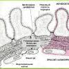
(Nephrotic syndrome requires attention from a qualified nephrologist. Only timely treatment can help cope with the disease. right and left respectively ). Each kidney is bean-shaped and covered with fibrous ( connective tissue ) and fat capsules ( shells ), which protect kidney tissue from damage. Inner sides ( medial sides ) kidneys are concave and are located next to the spine, external () – convex and facing the walls of the abdomen and lower back. On the medial side of each kidney there is a gate, through which vessels and nerves enter the kidneys.
The inside of the kidneys is made up of connective tissue ( interstitium) and the system of formation and excretion of urine. If you make a cut, you can see that the entire parenchyma of the kidney is not homogeneous. It includes the so-called medulla and cortex, which differ from each other in color, location and density. The cortex occupies the outer part of each kidney; it is relatively smaller in volume and density than the medulla, which is localized in the central part of the organ. The cortex is yellowish-red, the medulla is bluish-red. The medulla structurally consists of cone-shaped formations ( renal pyramids), the bases of which are directed towards the cortex. The apices of these pyramids are directed towards the hilum of the kidney.
The system of formation and excretion of urine, in fact, begins in the cortex, where a huge number of nephrons are located - the main functional units of the kidneys. Each nephron consists of a vascular glomerulus ( glomeruli), capsules ( Shumlyansky-Bowman) and tubules. The choroidal glomerulus is a system of branching and repeatedly intertwining small arterial vessels located in the capsule. The arterial capillaries in the capsule are surrounded by mesangium ( type of connective tissue). The capsule itself looks like a bowl ( in which the glomerulus is immersed) and consists of two leaves - outer and inner. The outer leaf is formed by connecting flat cells to each other, forming a single-layer membrane.
The structure of the inner leaf is a little more complex. It consists of three shells. The first membrane is the cells of the capillary walls ( endothelial cells) choroid glomerulus. The second membrane is the basement membrane, to which endothelial cells are attached. This membrane is located outside the capillaries ( and outside of endothelial cells). The third layer of the inner layer of the Shumlyansky-Bowman capsule is a layer of podocytes ( special epithelial cells). They are localized on the basement membrane on the reverse side of the endothelial cells. Podocytes have processes that cover the capillaries of the vascular glomerulus. All three membranes of the inner layer of the Shumlyansky-Bowman capsule make up the so-called glomerular filter ( barrier). Through it, filtration occurs in each nephron of the kidneys ( straining) blood ( entering the kidney via the renal artery), resulting in the cavity of the capsule itself ( which is located between its outer and inner leaves) primary urine is formed ( glomerular ultrafiltrate).
Primary urine is blood plasma freed from formed elements and large molecular compounds ( mostly proteins). To prevent the body from losing everything valuable ( for example, water, mineral salts, vitamins, amino acids, etc.) that is in the primary urine, it must pass through the tubular system - the last component of each of the nephrons. Reabsorption occurs in the tubules ( reverse suction) substances useful for the body from primary urine back into the blood. Filtration and reabsorption are the main functions of the renal nephrons. Most of the tubules are located in the renal medulla. After passing through the tubule system, primary urine gradually turns into secondary urine, which is excreted from the tubules ( and, strictly speaking, from the nephrons themselves) into the renal calyces, localized in the area of the apices of the renal pyramids. Connecting with each other, these calyces flow into the renal pelvis, through which the secondary ( final) urine penetrates further into the ureter and is excreted from the kidneys.
The arterial blood supply to the kidneys is provided by the renal arteries, which branch from the abdominal aorta. Venous blood from the kidneys flows through the renal veins. These veins then drain into the inferior vena cava. Lymphatic vessels deliver all the lymph to the lumbar lymph nodes. Innervation of the kidneys is provided through the branches of the renal plexus and nerves coming from the upper lumbar and lower thoracic nodes.
Causes and pathogenesis of nephrotic syndrome
Nephrotic syndrome is a set of clinical and laboratory abnormalities indicating a disorder of renal function. It is characterized by hypoproteinemia ( decreased protein levels in the blood), hypoalbuminemia ( decrease in blood albumin levels), proteinuria ( protein excretion in urine), edema and sometimes hyperlipidemia ( increased blood fat levels).Nephrotic syndrome is not an independent disease. It can occur in various diseases and pathological conditions. Moreover, it is not necessary that they ( these diseases and conditions) originally appeared in the kidneys. For example, nephrotic syndrome often occurs with blood diseases, diabetes mellitus, rheumatic diseases, systemic vasculitis ( pathologies associated with vascular inflammation) etc. In some cases, it, of course, can develop against the background of primary kidney diseases - acute glomerulonephritis, some genetic kidney diseases ( congenital nephrotic syndrome). In any case, the main mechanism for the appearance of nephrotic syndrome is damage to the glomerular filter in the renal nephrons, against the background of which the process of filtration of blood plasma in the kidneys is disrupted.
The most common causes of nephrotic syndrome are:
- congenital nephrotic syndrome;
- acute glomerulonephritis;
- tubulointerstitial nephritis;
- amyloidosis;
- diabetes;
- infectious diseases;
- rheumatic diseases;
- blood diseases;
- systemic vasculitis;
- venous thrombosis;
- allergic diseases;
- poisoning with toxic substances.
Congenital nephrotic syndrome
Congenital nephrotic syndrome ( VNS) is a nephrotic syndrome that appears in children in the first 3 months of life. In addition to the congenital, there is also the infantile ( infant) nephrotic syndrome, which first occurs in children 4 to 12 months of age. The causes of congenital and infantile nephrotic syndrome, as a rule, are various genetic disorders, less often infections and metabolic disorders. The most common genetic disorders that provoke the appearance of congenital nephrotic syndrome are mutations in the fibrocystin genes ( polycystic kidney disease), collagen type IV ( Alport syndrome), nephrine ( NPHS1 gene), podocin ( NPHS1 gene), WT1 gene ( Denis-Drash syndrome), PLCE1 gene ( impaired secretion of nephrin and podocin), LamB2 gene ( Pearson syndrome).All these genes are responsible for the proper development of kidney tissue during fetal embryogenesis, as well as for its functioning after the birth of a child. For example, a mutation in the NPHS1 gene ( Finnish type nephrotic syndrome) leads to disruption of the formation of nephrin protein in kidney cells, which serves as an important component of the gap spaces located between the podocyte stalks ( glomerular cells covering arterial capillaries entering Bowman's capsule). This is accompanied by destructuring of the capsule and impaired filtration of blood plasma, resulting in the development of nephrotic syndrome.
Congenital nephrotic syndrome can develop against the background of a variety of infectious diseases. This is often observed with intrauterine infections ( syphilis, cytomegalovirus infection, herpes, rubella, hepatitis B, toxoplasmosis, etc.), damaging various organs of the fetus ( including the kidneys) during pregnancy. Congenital nephrotic syndrome can also be caused by various metabolic disorders ( metabolic disorders), such as Fabry disease ( mutation in the a-galactosidase gene, leading to fat deposition in the glomerular region), congenital forms of hypothyroidism ( decreased thyroid function), hypoadrenocorticism ( adrenal gland dysfunction) and etc.
Acute glomerulonephritis
Acute glomerulonephritis is a kidney disease in which inflammation of a large number of glomeruli occurs in them ( glomerulus). The reason for its development is a disruption of the immune system, as a result of which it attacks its own kidney tissue. Acute glomerulonephritis most often appears after streptococcal pharyngitis ( inflammation of the pharyngeal mucosa). As the body fights this infection, pathogenic streptococci are partially destroyed. The remaining bacterial particles settle in the glomeruli. After a few weeks, the immune system can detect deposited streptococcal particles and initiate an immune response that causes inflammation of the glomeruli.Inflammatory changes in the glomeruli often lead to damage to their morphological structure and an increase in their permeability to various plasma components that normally do not penetrate into the primary urine ( for example, large molecular proteins, blood cells). Therefore, kidney damage in acute glomerulonephritis is often accompanied by signs characteristic of nephrotic syndrome ( proteinuria, hypoalbuminemia and edema). In addition to streptococcal infection, acute glomerulonephritis can also be caused by infective endocarditis, rubella, pneumonia, measles, malaria, schistosomiasis, certain medications, etc.
Tubulointerstitial nephritis
Tubulointerstitial nephritis is a pathology that develops as a result of inflammation of the interstitial ( intermediate) kidney tissue, as well as renal tubules. With this disease, the function of the renal glomeruli is practically not impaired ( up to the most advanced clinical stages of the disease), so nephrotic syndrome is not so typical for him. However, there are cases when it was still observed with tubulointerstitial nephritis. The main causes of this renal pathology are pyelonephritis, leptospirosis, acute tubular necrosis ( instantaneous death of the renal tubular epithelium), caused by drugs, toxins ( lead, cadmium), ischemia ( impaired blood supply to the kidneys), radiation, obstructive nephropathy ( a pathology in which the normal flow of urine through the urinary tract is disrupted), tuberculosis, etc.The occurrence of nephrotic syndrome in tubulointerstitial nephritis is associated with the spread of the inflammatory process from the renal tubules and interstitium ( intermediate kidney tissue) to glomeruli. The development of inflammation in the glomeruli is then accompanied by a violation of their filtration function and the penetration of excess amounts of protein from the blood plasma into the primary urine. In addition, inflammation in the glomeruli in tubulointerstitial nephritis may also be promoted by the same etiological factors that provoked the onset of the underlying disease. For example, if tubulointerstitial nephritis was caused by poisoning of the patient with salts of heavy metals ( lead, cadmium), then they can also serve as damaging agents in relation to the anatomical structures of the renal glomeruli.
Amyloidosis
Amyloidosis is a disease caused by a disorder of protein metabolism. It is characterized by the formation of a pathological protein in the body - amyloid, which over time settles in various tissues and organs ( liver, heart, kidneys, gastrointestinal tract, etc.), disrupting their basic functions. Today, many types of amyloidosis are known, which differ from each other depending on the origin of the amyloid protein. There are so-called genetically determined types of amyloidosis ( e.g. ATTR amyloidosis, Finnish type amyloidosis, AF amyloidosis), in which the appearance of amyloid is clearly associated with genetic mutations in certain proteins of the body. In addition to them, secondary forms of amyloidosis are known, associated with various diseases, for example, myeloma ( AL amyloidosis), Alzheimer's disease ( AB amyloidosis), chronic diseases ( AA amyloidosis), tumors ( AE amyloidosis) and etc.In all of the above types of amyloidosis, an abnormal amyloid protein is formed, deposited in the kidney tissue and gradually causing kidney failure. At the initial stages, amyloid deposition occurs in the zone of the basement membranes of the renal glomeruli and their mesangium ( tissue located between capillaries in nephron capsules). The constant sedimentation of new masses of amyloid in the glomeruli leads to disruption of their structure ( which is accompanied by nephrotic syndrome) and progressive replacement of normal elements ( capillaries, mesangium, podocytes, etc.), forming nephron capsules. Amyloid accumulation may also be observed in the renal interstitium ( intermediate kidney tissue), peritubular ( near the tubules) and perivascular tissue. Thus, in the process of amyloid deposition, mechanical replacement of normal kidney tissue with abnormal – amyloid – is observed, resulting in the development of nephrotic syndrome.
Diabetes
Diabetes mellitus is an endocrine disease that is associated with an absolute or relative deficiency of the hormone insulin, produced in the pancreas and regulating metabolism in the body. Insulin primarily regulates blood glucose levels ( Sahara) and lowers its level if it goes off scale. In diabetes mellitus, either an insufficient amount of this hormone is produced, or the action of insulin is simply ineffective on the target tissue ( that is, those tissues on which it should exert its direct effect). Therefore, diabetes mellitus is accompanied by an increase in blood glucose.Diabetes mellitus can cause the development of nephrotic syndrome. The fact is that with this endocrine disease due to hyperglycemia ( increased blood glucose levels) glycosylation occurs ( non-enzymatic addition of glucose to other chemical compounds) various proteins that make up the various structures of renal nephrons ( capillary walls, mesangium, basement membrane, etc.). This leads to an increase in the permeability of the glomerular filter for proteins, which is accompanied by the release of a significant amount of it into the primary urine and, as a consequence, the development of nephrotic syndrome.
The formation of nephrotic syndrome in diabetes mellitus is also facilitated by increased blood pressure, which disrupts intraglomerular blood flow ( blood pressure inside the glomeruli increases significantly, which creates additional stress on the walls of the intraglomerular capillaries). Since hyperglycemia is a constant phenomenon in diabetes mellitus, the glycosylation of proteins in the renal glomeruli gradually progresses, which creates the preconditions for the development of renal sclerosis ( ) and the appearance of renal failure. All pathological changes observed in the kidneys with diabetes are called diabetic nephropathy. It is one of the complications of diabetes mellitus and may not always occur in a patient suffering from this endocrine disease.
Infectious diseases
In some infectious diseases, the pathogen may be carried into the kidneys. This often happens with various bacterial ( tuberculosis, septic endocarditis, pneumonia, syphilis, abscesses, bronchiectasis, osteomyelitis, etc.), viral ( HIV infections, etc.) and fungal ( actinomycosis) pathologies. Penetration of pathogenic microorganisms ( viruses, bacteria, fungi) into the kidney tissue usually occurs through the blood ( hematogenously). Once in the kidneys, they damage their various tissue structures.Most often in such cases, the cells of the glomerular capillaries are affected ( endothelial cells), mesangium and podocytes ( glomerular cells lining arterial capillaries). Since these cells are part of the glomerular filter, their gradual death is accompanied by a violation of its permeability and increased release of proteins from the blood plasma into the primary urine. It is this mechanism that underlies the appearance of nephrotic syndrome in infectious diseases. Also, the inflammatory process that develops immediately at the site of damage to endothelial cells, mesangium and podocytes can play some role in its appearance during infections. With such inflammation, the basement membrane of the glomerular filter is often damaged, which only further intensifies the disorders that arose before.
Rheumatic diseases
Nephrotic syndrome can develop in many rheumatic diseases ( systemic) diseases ( for example, systemic lupus erythematosus, rheumatoid arthritis, systemic scleroderma, rheumatism, etc.). In systemic lupus erythematosus, the kidneys are often affected due to the deposition of immune complexes ( connection between an antibody and an antigen - a molecule foreign to the body) in the region of the renal glomeruli. The accumulation of such complexes leads to the development of an immunoinflammatory response from the body, damage to the anatomical structures of the glomeruli and the development of nephrotic syndrome.The appearance of this syndrome in rheumatoid arthritis, in most cases, is associated either with secondary renal amyloidosis, in which the renal glomeruli become clogged with an abnormal protein - amyloid, or with damage to glomerular tissue by antirheumatic drugs. In systemic scleroderma, nephrotic syndrome develops as a result of a disruption in the interaction of cells of the immune system with cells of other tissues ( vascular, connective, renal, etc.), which leads to the occurrence of inflammatory processes in the glomeruli and their sclerosis ( replacing them with connective non-functional tissue).
In fact, rheumatic diseases contribute to the development of inflammatory reactions in the glomeruli, which are accompanied by impaired permeability of the glomerular filter and the development of nephrotic syndrome. In fact, in such cases glomerulonephritis develops ( a disease in which diffuse inflammation of the renal glomeruli is observed), and this glomerulonephritis has a long course and is not acute, but chronic. Chronic glomerulonephritis can sometimes occur for a long time without any symptoms. The main danger of this disease is that over time it can lead to chronic renal failure. Therefore, all patients with rheumatic diseases should periodically monitor the functional state of their kidneys.
Blood diseases
Nephrotic syndrome can occur with certain blood diseases, for example, lymphogranulomatosis, myeloma, mixed cryoglobulinemia, thalassemia, sickle cell anemia, etc. With lymphogranulomatosis ( tumor developing from cells of the lymphoid system) the kidneys are rarely affected, but if this happens, then most likely this blood disease has been bothering the patient for a long time. Typically, kidney tissue damage is observed in stages 3 or 4 ( final stage) lymphogranulomatosis, in which not only the lymphatic system, but also many other organs are involved in the pathological process ( besides the kidneys), which is associated with the spread of tumor cells throughout the body. Once in the kidney tissue, such cells actively multiply and replace normal tissue with malignant tissue, as a result of which the internal structure of the kidneys is disrupted and nephrotic syndrome develops.For multiple myeloma ( malignant tumor of plasma cells - special blood cells) the so-called myeloma nephropathy develops, which is characterized by damage to intrarenal structures due to the penetration of abnormal myeloma proteins into them ( paraproteins), secreted by plasmacytoma cells ( myeloma tumor). Kidney damage in multiple myeloma is often accompanied by nephrotic syndrome, nephrosclerosis ( replacement of renal tissue with connective tissue) and renal failure. With mixed cryoglobulinemia, specific protein molecules, cryoglobulins, deposit on the walls of the vessels of the renal glomeruli, which initiate the local launch of inflammatory reactions ( via the complement system), which is accompanied by damage to the glomerular filter and the development of nephrotic syndrome in the patient. The appearance of cryoglobulins is caused by a disorder in the functioning of cells of the immune system, which occurs during certain viral infections ( hepatitis B, hepatitis C, cytomegalovirus infection, infectious mononucleosis, etc.).
Development of nephrotic syndrome in thalassemia ( genetic disease associated with impaired hemoglobin formation) is caused by damage to the glomeruli, against the background of periodic accumulation of iron in them, formed during the breakdown of pathological red blood cells. For sickle cell anemia ( a disease in which there is a disruption in the formation of normal hemoglobin) thrombosis often occurs in the kidneys ( blockages of blood vessels), due to the reduced resistance of red blood cells to destruction. Against the background of thrombosis in the glomeruli, blood flow is often disrupted, which often leads to hyperfiltration ( increased filtration) plasma through the glomerular filter and the appearance of nephrotic syndrome.
Systemic vasculitis
Systemic vasculitis is a group of diseases in which inflammation of the walls of blood vessels located in various tissues and organs is observed. The renal vessels are most often damaged in several systemic vasculitis ( polyarteritis nodosa, Henoch Schönlein purpura and Wegener's granulomatosis). The mechanism of origin of all three pathologies is associated with disruption of the immune system. For example, Henoch's Schonlein purpura occurs as a result of the deposition of immune complexes on the walls of blood vessels, as a result of which the complement system is activated, which triggers a local inflammatory reaction and promotes them ( vessel walls) damage. The origin of the immune complexes themselves has not yet been precisely established; it is assumed that they may be molecules remaining in the patient’s body after certain diseases ( allergies, streptococcal infection, mycoplasma infection, etc.).The mechanism of development of inflammation of the walls of renal vessels in polyarteritis nodosa is in general similar to that observed in Henoch Schönlein purpura, however, the occurrence of immune complexes in this pathology may also be associated with some other viruses ( for example, hepatitis B virus, hepatitis C virus, HIV infection, cytomegalovirus) or certain drugs ( bismuth preparations, antibiotics, sulfonamides, etc.). The pathogenetic mechanism of origin of Wegener's granulomatosis is even more complex. It includes not only precipitation ( deposition) on the walls of blood vessels of glomeruli of immune complexes, but also various disorders of interaction between cells of the immune system. Neutrophils play an important role in damage to the walls of blood vessels in this disease ( blood cells), which accumulate in the area of the renal glomeruli and produce various enzymes that have a damaging effect on them. Damage to blood vessels in all three types of systemic vasculitis leads to impaired permeability of the glomerular filter and the development of nephrotic syndrome.
Venous thrombosis
Nephrotic syndrome can be detected in patients with thrombosis of large venous vessels ( inferior vena cava, renal veins). Such thromboses are a common occurrence in diseases and conditions associated with blood clotting disorders ( taking oral contraceptives, pregnancy, congenital diseases of the blood coagulation system, etc.). In addition, thrombosis is periodically observed in heart failure, systemic vasculitis, tumor diseases ( when the venous vessels are compressed from the outside by malignant formations).With thrombosis in the venous vessels, blood flow is disrupted, which contributes to blood stagnation and retrograde ( the opposite) increased pressure in the renal arteries. An increase in pressure in the glomerular arterial system leads to dilation of the vessel walls and increased plasma filtration. Therefore, thrombosis of large venous vessels, such as the inferior vena cava, renal and other veins, is quite often associated with nephrotic syndrome. Other common causes of thrombosis in these veins include malignant kidney tumors, metastases, severe dehydration ( in children), aortic aneurysm, severe trauma, sepsis, peritonitis ( inflammation of the peritoneum), antiphospholipid syndrome, etc.
Allergic diseases
For some allergic pathologies ( for example, hay fever, allergies to food, cosmetics, allergic reactions that occur after insect bites, etc.) nephrotic syndrome can sometimes develop. The mechanism of its appearance is, in general, similar to that which occurs in rheumatoid diseases ( for example, systemic lupus erythematosus) and is caused by the accumulation of immune complexes on the walls of the vessels of the renal glomeruli, as a result of which their immunoinflammatory damage unfolds, leading to disruption of the normal permeability of the glomerular filter and hyperfiltration ( increased filtration) the liquid part of the blood through it. Unlike systemic lupus erythematosus, damage to the glomeruli in allergic diseases is most often temporary. Nephrotic syndrome immediately ( or after a short period of time) disappears after eliminating the allergen ( etiological factor that caused the allergy).Poisoning with toxic substances
Poisoning with toxic substances quite often leads to kidney damage and the development of nephrotic syndrome. This is often observed when various pesticides and toxic poisons enter the patient’s body ( ethylene glycol, oxalic acid, arsenic, acetic acid, chromium, lead, radioactive elements, copper sulfate, arsine, etc.). In rare cases, the use of certain medications ( for example, antiepileptic drugs, bismuth drugs, gold, mercury, antibiotics, anticoagulants, sulfonamides, vitamins, D-penicillamine) can also cause nephrotic syndrome.The mechanism of development of nephrotic syndrome itself in case of poisoning with toxic substances always depends on their type. For example, poisoning with substances containing mercury is accompanied by impaired cellular respiration of glomerular cells of the kidneys, which inevitably leads to their death. In case of poisoning with acetic acid, copper sulfate, arsine, which, by their nature, are hemolytic poisons ( that is, substances that destroy red blood cells), excessive deposition of free hemoglobin occurs ( released when red blood cells break down) in the area of glomerular filters, which leads to nephrotic syndrome.
Damage to kidney tissue by toxic substances is called toxic nephropathy. It can be acute or chronic. Acute toxic nephropathy usually leads to the development of acute glomerulonephritis ( inflammation of the glomeruli of the kidneys) and then to acute renal failure. Chronic nephropathy is accompanied by the appearance of chronic glomerulonephritis and chronic renal failure. Nephrotic syndrome in toxic nephropathy is caused precisely by acute or chronic glomerulonephritis.
Nephrotic syndrome is not an independent disease, but rather a combination of causes, signs and consequences of filter damage of various natures.
 Nephrotic syndrome is of two types - primary and secondary. The division into these types is due to the root cause of its development. Primary nephrotic syndrome is caused by kidney disease. In the secondary type, nephrotic syndrome appears due to the presence of pathologies in the patient that primarily affect other organs rather than the kidneys. There is also a classification of nephrotic syndrome depending on the degree of its response to treatment with hormonal drugs ( prednisone). In this classification, nephrotic syndrome is divided into steroid-resistant and steroid-sensitive types.
 Primary nephrotic syndrome
Primary nephrotic syndrome develops with acute or chronic glomerulonephritis ( inflammation of the glomeruli of the kidneys) and some genetic kidney diseases ( congenital nephrotic syndrome). Idiopathic nephrotic syndrome is also classified as primary nephrotic syndrome. In idiopathic nephrotic syndrome, the cause of kidney damage is unknown.Secondary nephrotic syndrome
Secondary nephrotic syndrome occurs with a variety of diseases of other organs and tissues, for example, allergies, systemic vasculitis, toxic substance poisoning, venous thrombosis, blood diseases, diabetes mellitus, amyloidosis, infectious and rheumatic diseases, tubulointerstitial nephritis. Damage to the kidney tissue in these pathologies is not always accompanied by nephrotic syndrome, and, in some cases, the kidneys are not affected at all, therefore it is believed that the development of this syndrome in these pathologies is secondary.Steroid-resistant nephrotic syndrome
Steroid-resistant nephrotic syndrome is a nephrotic syndrome that does not disappear in humans ( not in remission) after an eight-week course of steroid therapy with prednisone.Steroid-sensitive nephrotic syndrome
Steroid-sensitive nephrotic syndrome ( SCNS) is observed in those patients who have a positive response to treatment with prednisolone. Usually remission ( disappearance of symptoms and signs of the disease) This syndrome occurs after 2–4 weeks of steroid treatment. In some cases, the period may be extended to 4–8 weeks. Relapses ( reappearance of symptoms and signs of the disease) nephrotic syndrome may occur after it goes into remission during treatment, or may not appear at all.Steroid-sensitive nephrotic syndrome is divided into several types ( non-relapsing, infrequently relapsing, frequently relapsing, steroid dependent). With non-recurrent SSNS, relapses of the disease do not occur, remission of the pathology is very long. With infrequently recurrent SSNS, relapses occur less than twice every 6 months, and with frequently recurrent SSNS, relapses occur at least twice every six months. In steroid-dependent SSNS, the appearance of relapses is usually associated with cessation of the course of hormonal therapy with prednisolone; sometimes such relapses may be due to a reduction in the dose of the drug ( prednisone) taken during treatment.
Symptoms of nephrotic syndrome
 The only specific symptom of nephrotic syndrome is swelling. Other symptoms ( for example, nausea, vomiting, pain in the heart, weakness, decreased performance, shortness of breath, impaired growth and development, etc.), which may occur in a patient with this pathology, are classified as its nonspecific manifestations. These symptoms are additive. They may or may not occur with it. Swelling always serves as one of the mandatory clinical criteria for nephrotic syndrome.
 The difficulty in diagnosing nephrotic syndrome is that this syndrome is more laboratory than clinical, since most of its signs are determined using laboratory tests and are not recognized during a routine examination of the patient. Therefore, to establish the fact that a patient has nephrotic syndrome, it is not enough to simply detect swelling on his body. In such cases, he must prescribe the necessary laboratory tests.
Main symptoms observed in patients with nephrotic syndrome
| Symptom | The mechanism of appearance of this symptom | How does this symptom manifest itself? |
| Edema | In nephrotic syndrome, the patient's body loses huge amounts of protein through the kidneys. This protein comes into the urine from blood plasma. Such constant losses are accompanied by a decrease in the amount of proteins in the blood. One of the functions of plasma proteins is to maintain blood oncotic pressure. This pressure determines in which direction the intravascular fluid will move. The less proteins in plasma, the lower its oncotic pressure. If the oncotic pressure in the vessels is low, then the liquid does not stay in them for a long time and leaves them, then entering the intercellular spaces ( because the pressure is higher in them) various tissues rich in subcutaneous fat, resulting in the formation of edema. | The severity of edema usually correlates ( tied up) with a degree of proteinuria ( that is, the intensity of the patient’s loss of protein in the urine). Swelling, as a rule, first appears in the area of the face, eyelids, lower back, and genitals. Further, the swelling spreads to the arms and legs, less often to the torso. The rate of occurrence and distribution of edema may vary. They can develop instantly, literally overnight, or they can appear gradually over several days, weeks and even months. When palpated, the swollen skin is soft ( loose). If you press on it, then immediately after that a hole is formed at the site of the depression. Edema areas in nephrotic syndrome move easily. Swelling of the external genitalia makes urination difficult. |
| Heartbeat | In most patients with nephrotic syndrome, fluid accumulates not only externally ( under the skin), causing peripheral edema, but also in various cavities of the body. If fluid accumulation occurs in the pericardial cavity ( cardiac sac), then the patient experiences palpitations and pain in the heart area. Accumulation of fluid in the chest cavity ( hydrothorax) causes shortness of breath. Fluid entering the abdominal cavity contributes to the development of ascites and the appearance of nausea, vomiting, diarrhea and bloating. | Cardiac symptoms ( pain, palpitations), are slightly less common than from the gastrointestinal tract ( nausea, vomiting, diarrhea) and lungs ( dyspnea). The intensity of these symptoms is always individual and is largely determined by the degree of proteinuria. The sequence of appearance of all these symptoms may vary. Gastrointestinal symptoms usually appear after eating. Shortness of breath, pain in the heart and palpitations are present in patients both at rest and during physical activity. |
| Pain in the heart area | ||
| Dyspnea | ||
| Nausea | ||
| Vomit | ||
| Diarrhea | ||
| Tearing | The appearance of lacrimation is caused by swelling of the conjunctiva ( mucous membrane) eye. | Watery eyes are an intermittent symptom of nephrotic syndrome. |
| Disorders of growth and development | Impaired growth and development occurs as a result of various metabolic disorders that can be identified in nephrotic syndrome. For example, with this pathology the amount of microelements in the body decreases ( iron, zinc, cobalt) and macroelements ( calcium), playing a great role in the development and growth of various tissues ( bones, nervous system, skin, cartilage, etc.). | Impaired growth and development are quite clearly observed in children suffering from nephrotic syndrome for a long time. Such children often lag behind their peers in development, both in general physical and intellectual terms. |
| Tetany | In nephrotic syndrome due to calcium metabolism disorders ( there is very little of it in the blood) convulsive attacks may be observed ( tetany). Hypocalcemia ( decrease in blood calcium) occurs as a result of a metabolic disorder of metabolites ( products of exchange) vitamin D in the body, which is involved in the regulation of phosphorus-calcium metabolism. | Tetany does not occur in all patients. It is more often observed in children and is combined with signs of osteomalacia and osteoporosis ( slowdown in the growth and development of bones, their excessive fragility, skeletal deformation, development of fractures, the appearance of pain in bones and muscles, etc.). |
| Skin paleness | Pallor of the skin, headache, decreased ability to work and weakness appear with nephrotic syndrome due to the presence of anemia ( decrease in the amount of hemoglobin and red blood cells in the blood). | These symptoms are quite common in patients with nephrotic syndrome. They appear periodically or are constantly present. Their intensity is always individual. |
| Headache | ||
| Decreased ability to work | ||
| Weakness | ||
| Weight gain | Weight gain is caused by the accumulation of additional fluid in the subcutaneous fat and various cavities of the body ( chest, abdominal, etc.). | An increase in body weight occurs as free fluid accumulates in the tissues. The rate of its increase cannot be predicted, since it depends on many factors ( the patient’s nutrition, the presence of additional diseases, the severity of nephrotic syndrome, etc.). |
| Dystrophic changes in the skin and its appendages | Dystrophic changes in the skin and its appendages ( nails, hair) in nephrotic syndrome are caused by disturbances in the metabolism of microelements ( zinc, iron, cobalt, copper) and a decrease in their concentration in the blood. | The skin with nephrotic syndrome is dry, rough, and often peels off. You can see cracks on it. Hair and nails become brittle and dull. |
Signs of nephrotic syndrome
Nephrotic syndrome is characterized by the appearance of certain symptoms in the patient. Almost all of these signs relate to laboratory indicators, so they are determined by conducting special studies. The main changes observed in nephrotic syndrome can be identified using a complete blood count, biochemical blood test, examination of the hemostatic system and a general urinalysis. The main sign of nephrotic syndrome in a general urinalysis is massive proteinuria ( protein excretion in urine), in which the total protein content in the urine should exceed 3.0 - 3.5 grams per day. Such protein losses are one of the main criteria for nephrotic syndrome in adults ( in addition to edema, hypoproteinemia and hypoalbuminemia).In children, the degree of proteinuria is calculated based on their body weight. In nephrotic syndrome, the amount of protein lost by their kidneys should be more than 50 mg per kilogram of body weight per day. In addition to proteinuria, leukocyturia can be observed quite often in a general urinalysis in nephrotic syndrome ( the appearance of leukocytes in the urine) and hematuria ( the appearance of red blood cells in the urine). Hyperaminoaciduria is much less common ( ), glucosuria ( ), hyperphosphaturia ( ).
The main signs of nephrotic syndrome in a biochemical blood test are hypoproteinemia ( ), hypoalbuminemia ( decrease in albumin in the blood), hyperlipidemia ( ), and secondary - an increase in urea, a decrease in the amount of iron, zinc, cobalt, calcium in the blood. The occurrence of hypoproteinemia in nephrotic syndrome is caused not only by damage to the glomerular filter and direct loss of plasma protein in the urine, which is observed in all diseases that cause this syndrome. Part of the plasma protein in nephrotic syndrome is removed from the vascular bed and undergoes intense catabolism ( the process of decay) in various tissues. In addition, with this syndrome, a lot of proteins, as a result of the development of peripheral edema, tend to leave the vessels into the interstitial ( intercellular) textile.
The total protein content in the blood serum in nephrotic syndrome decreases to 25–40 g/l. Such changes indicate a fairly pronounced degree of hypoproteinemia. The mechanisms of development of hypoalbuminemia in nephrotic syndrome are similar to those that cause the occurrence of hypoproteinemia. The concentration of albumin in blood plasma with this pathology can drop to 8–35 g/l. Since albumins perform many important functions in the circulatory system ( for example, they transport hormones, drugs, minerals, bind various toxic metabolites and transfer them to the liver, etc.), then a drop in their concentration leads to metabolic disorders ( metabolism) throughout the body. Albumin also helps maintain blood oncotic pressure. Therefore, their decrease is accompanied by an even greater increase in peripheral edema.
Hyperlipidemia does not serve as a baseline ( optional) criterion for diagnosing nephrotic syndrome, but it is one of the main ( very frequent, but not always occurring) its signs. Hyperlipidemia is a general term for an increase in the amount of fat in a patient's blood, but at its core it includes abnormal changes in several parameters ( some of which include not only fats). In particular, with nephrotic syndrome there is an increase in the blood levels of total cholesterol, non-esterified fatty acids, triglycerides, and low-density lipoproteins ( LDL), apoprotein B.
The origin of hyperlipidemia in this syndrome is explained by several theories. The main one is the one based on the mechanism of hypoalbuminemia. The fact is that hypoalbuminemia stimulates the synthesis in the liver of an increased amount of blood plasma proteins and special transporters ( lipoproteins), which transport fats in the blood. Since the patient has nephrotic syndrome, most of the protein produced in the liver is lost through the kidneys ( while the increased amount of lipoproteins in the blood persists and gradually increases, which indirectly contributes to the accumulation of fats).
Another theory suggests that hyperlipidemia in nephrotic syndrome is partly due to impaired breakdown processes ( catabolism) fats, due to disruption of certain enzymes ( for example, lipoprotein lipases) responsible for this process. The presence of hyperlipidemia in nephrotic syndrome has a detrimental effect on the state of the cardiovascular system ( promotes the development of atherosclerosis) and worsens the prognosis of the syndrome itself, since the increased content of fats in the blood ultimately leads to their deposition in the structures of the renal glomeruli ( especially those that are damaged). The accumulation of fat in the glomeruli stimulates the development of inflammatory and sclerotic changes in them, which contribute to the strengthening of nephrotic syndrome.
Changes in the hemostatic system ( that is, the blood clotting system) in nephrotic syndrome affect its cellular and plasma-coagulation link. If we talk about changes in the cellular unit, then here, mainly, pathological changes are observed in platelets, which acquire an increased ability to adhesion ( adhesion to any surface, usually to the vascular endothelium) and to aggregation ( platelets sticking together). This is caused by an increase in the amount of fibrinogen in the blood plasma, as well as hyperlipidemia ( increased amount of fats in the blood), hypoalbuminemia ( decrease in the amount of albumin in the blood) and a decrease in the production of nitric oxide in vascular endothelial cells, which prevents excessive platelet aggregation.
In the plasma-coagulation link of hemostasis, in addition to increasing the fibrinogen already mentioned just above, the concentration of some coagulation factors in the blood also increases ( ). In addition to the functioning of the blood coagulation system in nephrotic syndrome, the functioning of the anticoagulation system, which normally balances with the coagulation system and controls its activity, is also disrupted. Disturbances in the anticoagulant system occur in the fibrinolysis system, which is responsible for the decomposition ( resorption) new blood clots in the vessels. These disorders are caused by the loss in urine of the main components of the fibrinolysis system ( mainly plasminogen). In nephrotic syndrome, the patient loses a sufficient amount of natural anticoagulants through the kidneys ( antithrombin III, protein S), preventing the formation of blood clots in blood vessels. Due to such extensive and serious changes in the hemostatic system, patients with nephrotic syndrome very often experience thrombosis of the deep veins of the legs, renal veins, coronary arteries and thromboembolic complications ( most often - pulmonary embolism - PE).
Diagnosis of nephrotic syndrome
 The diagnosis of nephrotic syndrome is made based on clinical results ( taking anamnesis, palpation, examination of the patient) and laboratory ( general, biochemical blood test, general urine test, study of the state of the hemostatic system) research. An important part of diagnosing this syndrome is establishing its cause ( since nephrotic syndrome is not an independent disease, but appears against the background of various other diseases). For this purpose, not only the methods used to diagnose nephrotic syndrome itself are prescribed, but also some other examinations, for example, radiation research methods ( ultrasound, computed tomography, magnetic resonance imaging), other laboratory tests ( immunological, genetic blood test, urine test according to Nechiporenko, according to Zimnitsky, etc.), kidney biopsy, electrocardiogram ( ECG), urography, scintigraphy, pulmonary radiography, etc. These additional studies help not only to detect the underlying pathology, but also to establish various complications of nephrotic syndrome.
 Methods for diagnosing nephrotic syndrome
| Method name | Methodology | What signs of nephrotic syndrome does this method reveal? |
| History taking | History ( anamnestic data) is information obtained by a doctor when questioning a patient during a medical consultation. The anamnestic data includes not only the patient’s complaints, but also other important information regarding the circumstances of the onset of the disease, its clinical course, as well as the living conditions of the patient himself. | In the process of collecting anamnesis, you can detect the main symptoms of nephrotic syndrome ( for example, swelling, nausea, vomiting, pain in the heart, weakness, decreased performance, shortness of breath, impaired growth and development, etc.), the sequence of their appearance and intensity. An important step in collecting anamnesis is to establish whether the patient has kidney or pancreas diseases ( diabetes mellitus), blood vessels, rheumatic diseases, and the use of various toxic substances that could contribute to the development of nephrotic syndrome. |
| Patient examination | The patient is examined by a doctor during a consultation. The essence of the method is a visual examination of the external integument of the patient’s body to identify abnormal changes. | In nephrotic syndrome, changes are most often found in the skin and its derivatives ( hair, nails), muscular system, joints. The skin of patients is usually swollen, with dystrophic changes ( flaky, cracked, pale, dry), hair and nails are brittle and dull. After swelling subsides, muscle atrophy can be detected ( decrease in muscle mass). Some joints may become enlarged due to fluid accumulation in them. |
| Palpation | Palpation ( palpation) the doctor examines certain areas of the patient’s body during his examination. | The main signs that can be detected by palpation are hepatomegaly ( liver enlargement) and swelling. Edema is the main clinical sign of nephrotic syndrome. Some swelling may not be detected during normal visual examination. In such cases, palpation helps. The doctor needs it to clarify the nature of the edema. Edema in nephrotic syndrome is soft, easily moved and has a dough-like consistency. |
| General blood analysis | Blood for general and biochemical blood tests is taken from the ulnar vein, in the morning, on an empty stomach. Then it is delivered to the laboratory, where it is analyzed in special biochemical and hematological analyzers. | A general blood test for nephrotic syndrome can reveal anemia ( decrease in the number of red blood cells and hemoglobin), increase in ESR ( erythrocyte sedimentation rate), eosinophilia ( increased levels of eosinophils in the blood), leukocytosis ( increase in white blood cell count) and lymphocytosis ( increase in the number of lymphocytes). |
| Blood chemistry | A biochemical blood test helps detect some changes in the patient’s biochemical composition of the blood, for example, hypoproteinemia ( decrease in total protein in the blood), hypoalbuminemia ( decrease in albumin in the blood), an increase in the concentration of cholesterol, non-esterified fatty acids, triglycerides, low-density lipoproteins ( LDL), apoprotein B, urea, creatinine, decreased amount of iron, zinc, cobalt, calcium. | |
| General urine analysis | Urine collection is carried out in the morning. Urine should be collected in sterile 200 ml plastic containers. The collected urine must then be delivered to the laboratory, where it will be biochemically and microscopically analyzed. | The main sign of nephrotic syndrome in a general urinalysis in adults is severe proteinuria ( protein excretion in urine), in which the total protein content in the urine exceeds 3.0 - 3.5 grams per day. In children, proteinuria, characteristic of nephrotic syndrome, should reach 50 mg/kg body weight/day or higher. Sometimes leukocyturia occurs with this syndrome ( the appearance of leukocytes in the urine), hematuria ( the appearance of red blood cells in the urine), hyperaminoaciduria ( excretion of increased amounts of amino acids in urine), glucosuria ( increased levels of glucose in urine), hyperphosphaturia ( excretion of increased amounts of phosphate in the urine). |
| Study of the hemostasis system | To study the hemostasis system, venous blood is taken on an empty stomach from the ulnar vein in the morning. After this, the blood is placed in special devices ( coagulometers and aggregometers), which calculate the main indicators of the hemostasis system. | In nephrotic syndrome, an increase in platelet adhesion and aggregation, hyperfibrinogenemia ( increase in the amount of fibrinogen in the blood), increased concentrations of some coagulation factors ( II, V, VII, VIII, XIII, von Willebrand factor), decrease in plasminogen, antithrombin III and protein S. Thrombocytosis ( increase in platelet count) is rarely detected in nephrotic syndrome. |
Treatment of nephrotic syndrome
 Treatment of nephrotic syndrome should be carried out in three main areas. The underlying pathology should be treated first ( for example, allergies, diabetes, infectious disease, etc.), which provoked the appearance of this syndrome. The dosages of all drugs and their choice, in such cases, depend on the disease that caused the nephrotic syndrome, its severity, the severity of the syndrome itself, the general condition of the patient, the presence of impaired function of various organs in the patient ( heart, kidneys, liver, intestines, etc.), his age, etc.
 The second direction of treatment is symptomatic and pathogenetic therapy of the syndrome itself, which is aimed at eliminating edema and reducing proteinuria ( protein excretion in urine), restoration of lipid balance and maintenance of normal functioning of the hemostatic system.
To reduce edema, diuretics are prescribed ( furosemide, veroshpiron). Sometimes diuretics ( diuretics) are prescribed together with dextran preparations ( reopolyglucin and reogluman), which improve microcirculation and are plasma substitutes. For very severe edema, salt-free albumin is prescribed ( to correct the total protein level in the blood plasma). To reduce protein excretion in the urine, glucocorticoids are prescribed ( prednisolone, methylprednisolone), cytostatics ( azathioprine, cyclosporine, cyclophosphamide, chlorobutine, mycophenolate mofetil, etc.), ACE inhibitors ( ramipril, enalapril, lisinopril). Glucocorticoids and cytostatics have an immunosuppressive effect on the patient’s immune system, due to which they help reduce inflammatory processes in the renal glomeruli and, thereby, reduce proteinuria.
The antiproteinuric effect of ACE inhibitors is that these drugs help reduce intraglomerular pressure in the capillaries of the glomeruli, and also reduce the permeability of the glomerular filter for large proteins. In addition, ACE inhibitors are used for nephrotic syndrome in cases where the patient's systemic blood pressure increases. To restore lipid balance, doctors usually prescribe HMG-CoA reductase inhibitors ( lovastatin), fibrates ( fenofibrate, gemfibrozil), probucol, nicotinic acid. Anticoagulants ( heparin) and antiplatelet agents ( dipyridamole, aspirin) are necessary for such patients to maintain the normal functioning of the hemostatic system and prevent thrombosis.
The third direction of treatment of nephrotic syndrome is therapy aimed at preventing and eliminating its complications ( nephrotic crisis, thrombosis, infectious diseases, acute renal failure, allergies, cerebral and retinal edema). The choice of drugs and their dosage in this direction depends on many factors ( the patient’s condition, the severity of nephrotic syndrome, the body’s response to the ongoing etiotropic therapy, the presence of diseases of other organs in the patient, etc.), determining the possibility of developing a particular complication.
Diet for nephrotic syndrome
 In case of nephrotic syndrome, it is necessary to limit the intake of table salt to 3 grams per day. This is necessary then to reduce the amount of sodium chloride entering the body, which is one of the factors causing the development of edema in nephrotic syndrome. It is advisable to exclude all salty foods from the diet ( pickles, marinades, seasonings, fish, etc.). It is especially important not to eat salty foods when swelling increases. Despite the fact that with this syndrome there is a significant loss of protein in the urine, the consumption of significant amounts of protein or intravenous administration of proteins does not improve the condition of such patients, so it is recommended to take a low-protein diet.
 The daily protein intake for such patients should be calculated based on their body weight ( 0.8 – 1.0 g/kg). In addition to salt and protein, all patients with nephrotic syndrome should also reduce their fat intake ( up to 70 – 85 g) per day. This measure allows you to level the intensity of hyperlipidemia ( a condition associated with increased amounts of fat in the blood), which quite often occurs in patients with nephrotic syndrome and serves as a risk factor for the development of atherosclerosis.
Foods you can and cannot eat if you have nephrotic syndrome
| Can be consumed | Cannot be consumed |
|
|

How long can nephrotic syndrome last?
 It is quite difficult to say exactly how long nephrotic syndrome can last, since its course and prognosis depend on many different factors ( the age of the patient, the nature of the kidney damage, the type of underlying disease, its duration, the adequacy of the medications used, the presence of complications, etc.). One of the important clinical prognostic indicators of nephrotic syndrome is the level of its response to hormonal and immunosuppressive therapy. If a patient develops steroid-resistant nephrotic syndrome, the prognosis of the disease is unfavorable. If the same activity is maintained, nephrotic syndrome will reach the terminal stage of chronic renal failure in 5–10 years.
 In steroid-sensitive nephrotic syndrome, remission ( transition of pathology to an inactive form), in most cases, observed after 2–8 weeks of intensive treatment. The only drawback of this form of nephrotic syndrome is that after entering remission, it may reappear, that is, a relapse may occur ( re-exacerbation). Predicting the risk of recurrence of steroid-sensitive nephrotic syndrome is as difficult as predicting the total duration of this syndrome in a patient.
Is nephrotic syndrome treated with folk remedies?
 Nephrotic syndrome is not recommended to be treated with folk remedies, due to their terribly low effectiveness. In addition, these drugs are not able to eliminate most of the main pathologies that contribute to the appearance of nephrotic syndrome. It is also worth recalling that the patient is most often unable to independently identify the presence of nephrotic syndrome in him or his loved ones, since for those close to him this syndrome is more laboratory than clinical. Peripheral edema is observed not only with nephrotic syndrome, but also with other diseases, for example, heart failure, liver cirrhosis, Parhon syndrome, intestinal amyloidosis, liver tumors, Crohn's disease, etc. Therefore, when edematous syndrome occurs, you should not immediately think about that the patient has developed nephrotic syndrome.
 Edema is only one of the main criteria for diagnosing this syndrome. Without laboratory tests, it is impossible to say for sure whether a patient has it or not. If the patient knows for sure that he has nephrotic syndrome and still wants to try some folk remedies, then in this case he needs to consult with his doctor before using them, since many folk remedies have side effects, which can be unfavorable affect the course of drug therapy.
What is the difference between nephritic and nephrotic syndromes?
 Nephritic syndrome, like nephrotic syndrome, is not a separate disease. It appears when the glomeruli become inflamed. The inflammatory process in nephritic syndrome is usually caused by damage to the glomerular structures by the patient's immune system. In nephritic syndrome, unlike nephrotic syndrome, the patient must have arterial hypertension ( high blood pressure) and urinary syndrome ( the appearance in the urine of red blood cells, protein in quantities less than 3.0 - 3.5 g/l, leukocytes and casts). Edema may also occur with nephritic syndrome. However, they are, for the most part, barely noticeable and are localized mainly on the face, especially in the eyelid area. Sometimes, with nephritic syndrome, the phalanges of the fingers and forearms of both hands become severely swollen. The development of edema in this syndrome is more due to sodium retention in the body than to a lack of proteins in the blood ( severe deficiency of proteins in the blood in nephritic syndrome, as a rule, is not observed), as occurs in nephrotic syndrome.
 What are the possible complications of nephrotic syndrome?
 The occurrence of complications in nephrotic syndrome depends on many factors ( the type of underlying pathology that caused this syndrome, its severity, the presence of additional diseases in the patient, the patient’s age, etc.). Most of these complications are quite serious and can threaten the patient's life, so it is very important to be aware of the possibility of their occurrence and prevent their development. In the vast majority of cases, complications of nephrotic syndrome appear with systemic lupus erythematosus, systemic vasculitis, renal amyloidosis, and diabetes mellitus.
 The main complications of nephrotic syndrome are:
- infections;
- swelling of the retina and brain;
- acute renal failure;
- nephrotic crisis;
- allergy;
- vascular disorders.
With nephrotic syndrome, patients often experience various infectious complications. This is due to a decrease in general resistance ( resistance) of their body against the background of constant losses in urine of protein and the gamma globulin fraction ( antibodies). In addition, when treating this pathology, doctors often prescribe steroidal anti-inflammatory drugs ( glucocorticoids), one of the side effects of which is immunosuppression ( suppression of immune system activity). Nephrotic syndrome usually involves a variety of bacterial infections, such as pneumonia ( pneumonia), pleurisy ( inflammation of the pleura), pleural empyema ( accumulation of pus in the pleural cavity), peritonitis ( inflammation of the peritoneum), sepsis, erysipelas of the skin. Sometimes viral and fungal infections can occur.
Swelling of the retina and brain
In some cases, nephrotic syndrome can cause vision problems. This is due to swelling of the retina. The retina is one of the layers of the eye in which the photoreceptor cells responsible for vision are located. The more pronounced the retinal edema, the more impaired the visual function of the eyes. Prolonged swelling can lead to vision loss, so it is very important to detect and eliminate it in time. Retinal edema in nephrotic syndrome is a temporary phenomenon; it immediately disappears when the required level of albumin in the blood is replenished and edema in other parts of the body disappears. Swelling of brain tissue is a very serious complication of nephrotic syndrome. Such swelling appears very rarely and, as a rule, occurs with massive swelling of the body. Its main symptoms are lethargy and severe lethargy of the patient. In such cases, mandatory medical intervention is required, since cerebral edema can develop into cerebral coma.
Acute renal failure
Acute renal failure ( surge arrester) is a pathology in which the kidneys suddenly stop performing their main functions ( carry out filtration and reabsorption) in full. AKI is not common in nephrotic syndrome. The causes of its appearance are usually renal vein thrombosis, sepsis, hypovolemic shock ( syndrome caused by a decrease in circulating blood volume), some medications ( non-steroidal anti-inflammatory drugs, diuretics). This complication is mainly observed in children or the elderly. Its main symptoms are oliguria ( decrease in the volume of urine excreted during the day), tachycardia ( increased heart rate), weakness, yellowing of the skin, increased blood pressure, nausea, abdominal pain, hyperkalemia ( increase in blood potassium), shortness of breath, increased blood concentrations of creatinine and urea.
Nephrotic crisis
Nephrotic crisis is one of the most severe complications of nephrotic syndrome, which sometimes leads to death. Nephrotic crisis is a situation in which there is a sharp deterioration in the general condition of patients with severe nephrotic syndrome, characterized by widespread edema of various parts of the body, massive effusions in the cavity ( chest, abdominal), significant hypoalbuminemia ( at which the concentration of albumin in the blood plasma drops to 10 g/l) and hypovolemia ( when the total volume of circulating blood decreases by 50–60% below normal). In a significant number of cases, this crisis develops spontaneously and unexpectedly.
The main symptoms of nephrotic crisis are severe abdominal pain, migrating erysipelas-like erythema ( redness) on the skin, changes in blood pressure ( it can increase or decrease), increased body temperature, leukocytosis ( increase in the number of leukocytes in the blood), leukocyturia ( presence of leukocytes in urine). Most often, nephrotic crisis appears with nephrotic syndrome caused by chronic glomerulonephritis, diabetes mellitus, lupus nephritis ( kidney damage due to systemic lupus erythematosus).
Provoking factors that contribute to its occurrence are inadequate therapy with diuretics ( in which there is a rapid and significant decrease in the total volume of circulating blood), as well as diarrhea, some infectious complications of nephrotic syndrome ( intestinal infections, pneumonia, skin infections, etc.). The final stage of the nephrotic crisis is hypovolemic shock - an abnormal condition in which there is a significant decrease in the amount of fluid circulating in the vessels, which leads to insufficient blood supply to tissues and organs and the development of multiple organ dysfunction ( dysfunction of various organs) due to a lack of oxygen that threatens the patient's life.
Allergy
In nephrotic syndrome, some patients may experience hypersensitivity ( increased sensitivity) to certain medications ( most often - to antibiotics) and food ( for example, chocolate, citrus fruits, milk, eggs, red fish, etc.). Allergic complications of this syndrome manifest themselves in the form of rashes of various types and locations. They may be similar to the rashes observed with erysipelas, psoriasis, lichen planus, eczematous dermatitis.
Vascular disorders
Vascular disorders in nephrotic syndrome arise due to various disorders in the hemostatic system ( increased platelet aggregation and adhesion, increased concentrations of fibrinogen, some blood clotting factors, decreased plasminogen levels, etc.) and the presence of severe hyperlipidemia ( increase in the amount of fats in the blood). The most common vascular disorders in this pathology are thrombosis of the veins of the lower extremities, renal, coronary ( heart) and cerebral ( brain) arteries and pulmonary embolism ( TELA). Thrombosis of the heart vessels often leads to the development of coronary heart disease and myocardial infarction.
Hyperlipidemia does not directly contribute to the development of thrombosis. In this condition, the walls of blood vessels change, lipids are deposited in them ( mainly cholesterol), which causes the appearance of atherosclerotic plaques that interfere with normal blood flow. Atherosclerotic changes underlie the development of cerebral and kidney strokes, coronary heart disease and myocardial infarction in such patients.
The kidneys take an active part in many vital processes of the body. The main task is to cleanse the blood of metabolic products and harmful impurities. Any violation in their work will entail a number of negative consequences. If a person has swelling on the face, legs, or arms, this is a signal to urgently undergo a kidney examination. Due to a decrease in the organ’s ability to filter fluids, nephrosis develops, another name is nephrotic syndrome. It contains a whole complex of various symptoms characteristic of a number of kidney diseases.
Overview of Nephrotic Kidney Syndrome
According to statistics, this pathology is diagnosed in 20% of cases of all kidney diseases. Most often the disease is detected in children two to five years old. In women and men, it is diagnosed between the ages of 20 and 40 years. Rarely affects the elderly and infants.
Nephrotic syndrome is usually described as a set of disorders in the functioning of the kidneys. But sometimes pathology acts as a separate nosological form. It is expressed in a low level of albumin (hypoalbuminemia), the release of protein into urine (proteinuria), and a decrease in it in the blood. But there is more fat.
Comes in three forms:
- Persistent syndrome is a common form, diagnosed in half of the cases. The disease is very sluggish, but progresses continuously. It is almost impossible to maintain a state of remission, which leads the patient to kidney failure within 8–10 years.
- Recurrent syndrome - occurs in approximately 20% of cases. It is characterized by a constant change of attenuation and exacerbation. In turn, recovery can occur either as a result of drug treatment or spontaneously. The latter option occurs infrequently and, as a rule, in children. This disease may have a period of rest for a long period (up to 10 years).
- Progressive syndrome - the disease progresses quickly and aggressively. Within a couple of years it leads to a chronic form of renal failure.
In medicine, nephrosis is classified as primary, secondary, idiopathic. Primary nephrotic syndrome develops due to kidney pathologies. It can be acquired or hereditary. The secondary cause is other inflammatory processes in the human body. But the mechanism of development of idiopathic nephrosis usually remains a mystery. This type is most often found in children.
If you look at how the symptoms of nephrotic syndrome manifest themselves, then they can distinguish an acute form of the disease, with pronounced symptoms, or a chronic one. The latter is characterized by alternating exacerbations and remissions. Occurs in adults who underwent poor or partial kidney treatment in childhood.
Doctors distinguish two more groups of nephrosis based on how the human body responds to hormonal therapy: 
- hormone-resistant nephrosis - that is, unresponsive to hormonal drugs, and therefore treatment is carried out with cytostatics that suppress cell growth and reproduction;
- hormone-sensitive nephrosis - responding to therapy with hormonal medications.
Causes of nephrotic syndrome development
The causes of the syndrome are varied and different for each type. Primary renal nephrosis may develop due to glomerulonephritis, hypernephroma (malignant tumor), nephropathy of pregnancy, or amyloidosis.
The following diseases cause the appearance of secondary nephrotic syndrome: periarteritis, diabetes mellitus, allergies, oncology, hemorrhagic vasculitis, lymphogranulomatosis, venous thrombosis, systemic connective tissue pathologies, some infections, prolonged septic endocarditis, HIV, hepatitis B, C. Secondary nephrosis can be provoked by drugs drugs: anticonvulsants, antibiotics, anti-inflammatory drugs or poisoning of the body with heavy metals (for example, lead, mercury, cadmium).

Vein thrombosis
To get rid of nephrotic syndrome, it is necessary to eliminate the cause of its occurrence. But this rule only applies if the source of the problem is a reaction to medication or a treatable disease. For example, for patients with glomerulonephritis without any particular complications, a favorable outcome of nephrosis occurs in more than 90%.
But when nephrosis becomes a consequence of an incurable disease progressing in the body, such as HIV, it is almost impossible to eliminate the syndrome. It becomes chronic with frequent relapses occurring in an acute form. The situation is the same with diseases that affect the entire body (diabetes, lupus). Therapy aimed at eliminating the main symptoms does not bring much results and over time the patient develops renal failure.
Most often, immune changes lead to nephrotic syndrome. Antigens circulating in the bloodstream prompt the human defense mechanism to produce antibodies to eliminate foreign particles.
Symptoms of nephrotic syndrome
Although there are many factors influencing the development of nephrotic syndrome, the symptoms of the pathological process are quite similar. The main sign indicating illness is swelling. At the beginning of the disease it is noticeable only on the face, especially in the eye area, then it appears in the lumbar region and genitals. Later, fluid accumulates in the intercostal space and around the lungs, peritoneum, cardiac sac, and subcutaneous tissue. If you press on the swelling, it will be soft. Quite mobile, “flows” when changing body position.

Swelling on the face
Due to the minimal content of nutrients, the patient’s skin peels, cracks, dries out, and oozes.
The accumulation of fluid in the heart sac (pericardium) leads to deterioration in the functioning of the organ. Pain and palpitations occur. Shortness of breath, even if the person is at rest, becomes a harbinger of hydrothorax (transudate in the chest). And nausea, indigestion, bloating, and vomiting appear after swelling of the abdominal cavity.
There are a number of other symptoms of nephrotic syndrome, of which you need to present:
- lethargy;
- weakness;
- decreased mobility;
- headache;
- pale skin;
- weight gain;
- decreased blood pressure, sometimes to the point of shock;
- loss of appetite;
- pulling, painful sensations in the abdomen and lumbar region;
- decrease in the amount of urine excreted from the body;
- dry mouth, thirst;
- increased blood cholesterol levels;
- muscle pain, cramps.

Convulsions
Signs of nephrotic syndrome appear and develop at a slow pace, but in the acute form of kidney nephrosis, the symptoms are violent and rapid.
Prolonged nephrotic syndrome has a bad effect on the mental and physical development of young children. Sick children usually lag behind their peers. This occurs due to disturbances in the body's metabolic processes. The level of macro- and microelements that affect the growth and development of bone, nerve, cartilage and other tissues decreases.
Diagnosis of kidney nephrosis
To make the correct diagnosis and choose treatment, the doctor conducts a detailed survey and examination of the patient. The following information is important for the medical history:
- general condition of the patient;
- risk factors;
- presence of infectious diseases;
- presence of systemic diseases;
- whether there were kidney pathologies in the family;
- when the first swelling is detected;
- whether the patient previously suffered from nephrosis (if so, what studies and treatment were performed).
The doctor conducts a visual examination of the patient's body. With nephrotic syndrome, there is a coating of the tongue, skin lesions, and an enlarged abdomen and liver. The skin feels cold and dry to the touch. If the liver is affected, a characteristic yellow tint appears. Swelling is more noticeable during the day and subsides slightly at night. In a patient with nephrosis, the nails peel off, become brittle, and the condition of the hair worsens. It is especially noticeable in women - they become dull and split.
Swelling of large joints (elbows, knees) is observed due to the accumulation of fluid in them. When tapping with the edge of the palm in the area of the renal projection, the patient feels pain (Pasternatsky's symptom).
A complete picture can only be obtained through a comprehensive clinical, instrumental and laboratory examination of the patient:
- General urine analysis. Helps evaluate sediment, chemical, physical state of urine. The following indicators are characteristic of nephrosis: the presence of red blood cells and protein in the urine, cylindruria. Mucus, fats, blood, and microbes may be present, making it cloudy.
- General blood analysis. Finger sampling is performed in the morning on an empty stomach. A study for nephrotic syndrome will show a high rate of leukocytes, platelets, low hemoglobin and red blood cells, and a high ESR. In this case, thrombocytosis is observed infrequently and is more likely a side effect of taking medications.
- Immunological analysis. Allows you to assess the state of the patient’s defense system and the presence of antibodies in the blood. The material is collected from a vein in the morning. Before this, you should not be very tired, drink, smoke, or eat.
- Blood chemistry. About 20 ml of venous blood will be required. Take it in the morning before meals. Shows disturbances in cholesterol and protein metabolism, kidney function. Albumin and protein decrease, the level of alpha globulins, cholesterol, and triglycerides increases. There may be signs of inflammation in the body if nephrosis occurs due to a chronic kidney infection.
- Bacterial examination of urine. Urine is usually sterile. But, if microbes are detected, then an analysis is prescribed in order to have a complete picture of the harmful small organisms. Urine is collected in a clean container after a thorough toilet. This will help prevent bacteria from entering from outside.
- For nephrosis, tests according to Nechiporenko, Zimnitsky and Reberg-Tareev are often prescribed. Among instrumental diagnostics, renal ultrasound, biopsy, dynamic scintigraphy, descending internal urography, cardiac electrocardiogram and lung x-ray are recommended. If there is a suspicion of nephrotic syndrome in a baby, then you should definitely consult a pediatrician and pediatric nephrologist.
Treatment of kidney nephrosis
Treatment of nephrotic syndrome is carried out in an inpatient setting under the guidance of a doctor. First, the underlying disease that caused the development of nephrosis is eliminated. Based on the general condition, severity, age and functional disorder of other organs, the doctor prescribes medications and determines the dosage.
Then the disease itself is treated, aimed at eliminating the symptoms. If nephrotic syndrome causes complications, then additional drugs are used to combat them.
During treatment, it is important to lead a calm lifestyle and not physically overwork the body, so that increased blood clotting does not provoke thrombosis.
Monitor the amount of fluid supplied. Its volume should not greatly exceed the amount of urine excreted from the body. Otherwise, swelling will only increase, which will not help get rid of kidney nephrosis at all.
Drug therapy for nephrotic syndrome
For nephrotic syndrome, the following drugs are prescribed: 
- diuretics – diuretic medications that help relieve swelling;
- cytostatics – control the growth of pathogenic cells;
- antibiotics - the dosage is determined by the attending physician and differs significantly for adults and children;
- glucocorticosteroids – have an anti-inflammatory, anti-edematous effect, reduce allergic manifestations;
- immunosuppressants - reduce the body's defenses so that it does not attack its own kidneys.
Additionally, to eliminate nephrotic syndrome, fresh frozen plasma or a plasma substitute, albumin, is prescribed. Typically, the full course of kidney treatment takes about eight weeks.
Traditional medicine for nephrosis
It is impossible to independently diagnose the syndrome at home. The appearance of edema does not always indicate the presence of nephrosis. Laboratory tests will be required, so at the slightest suspicion it is better to go to the clinic and undergo a comprehensive examination.
Folk remedies for the treatment of nephrotic syndrome have very low effectiveness and do not completely eliminate the causes and symptoms of the disease. Infusions and decoctions of medicinal herbs, which have anti-inflammatory and diuretic effects, should be used as an additional therapeutic agent. Before use, you should consult with your doctor so as not to aggravate the situation and prevent complications. 
Other treatments for nephrosis
Along with drug treatment of nephrotic syndrome, adherence to a strict salt-free diet is recommended. You are allowed to consume no more than three grams of salt per day. Any smoked meats, canned food, pickles, and seasonings are excluded from the diet. Protein foods are limited (up to 3 g per kilogram of body weight), fats.
Among the products allowed for nephrosis:
- butter and vegetable oil;
- eggs;
- low-fat dairy products;
- porridge;
- broths;
- lean meat and fish.
Those with a sweet tooth can treat themselves to berries, fruits, natural juices, honey, and jam.
Do not eat if you have nephrosis: 
- fatty dairy products;
- fatty fish and meat;
- raisins, nuts;
- ice cream;
- legumes;
- vegetables;
- chocolate;
- alcohol;
- coffee;
- cocoa;
- sparkling water and drinks.
Limit your intake of foods high in sodium and increase those rich in vitamins and potassium.
Preventive measures for nephrosis
To avoid the development of nephrotic syndrome, it is necessary to lead a normal lifestyle and control the diet not only during the period of exacerbation of the pathology, but also constantly. Closely monitor your health, especially if you have a family history of kidney disease or others that increase the risk of developing nephrosis. Eliminate infectious diseases in a timely manner, do not refuse recommended hospitalization and drug therapy. Use medications with nephrotoxic and allergic effects with caution.
Complications of nephrotic syndrome
If nephrotic syndrome is untimely or poorly treated, the pathology can be complicated by other diseases:

Pulmonary edema
- thrombosis;
- atherosclerosis;
- cerebral edema;
- myocardial infarction;
- nephrotic crisis.
A decrease in the human body’s immune defense contributes to the development of infectious diseases.
Nephrotic syndrome is a very complex pathology and it is rare to achieve complete recovery of the kidneys and stable remission. A positive outcome of the disease depends on many criteria: the patient’s age, degree of neglect, complications. Good prognosis for children, but subject to complete and high-quality medical treatment.
Nephrotic syndrome is a disease characterized by severe proteinuria, edema, hyperlipidemia, hypoalbuminemia, increased blood clotting, and lipiduria. Due to various immune disorders, metabolic disorders, toxic effects, and degenerative processes in the body, changes occur in the walls of the glomerular capillaries, causing excessive filtration of plasma proteins. Thus, acute nephrotic syndrome is a consequence of pathological conditions leading to increased permeability of the glomerular capillary wall.
The main causes of the development of nephrotic syndrome
Immunological mechanisms play a huge role in the development of nephrotic syndrome. The main reasons for the development of primary neurotic syndrome are:
- mesangial proliferative glomerulonephritis;
- membranous glomerulonephritis (idiopathic nephrotic syndrome in adults);
- membranous proliferative glomerulonephritis;
- lipoid nephrosis (minimal change nephropathy, idiopathic nephrotic syndrome in children);
- focal segmental glomerulosclerosis.
Secondary nephrotic syndrome develops as a result of:
- infectious diseases (leprosy, secondary syphilis, infective endocarditis, hepatitis B, etc.);
- toxic and medicinal effects (heavy metals, non-steroidal anti-inflammatory drugs, penicillamine, antitoxins, captopril, etc.);
- systemic connective tissue diseases;
- family hereditary diseases;
- other immune diseases;
- amyloidosis;
- rheumatoid arthritis;
- sickle cell anemia;
- diabetes mellitus;
- systemic lupus erythematosus;
- sarcoidosis;
- tumors (leukemia, lymphoma, melanoma, carcinoma, lymphogranulomatosis);
- Henoch-Schönlein disease.
It should be noted that nephrotic syndrome occurs approximately four times more often in children than in adults.
Symptoms of nephrotic syndrome
The main clinical symptom of nephrotic syndrome is edema. Edema can develop gradually, but in some patients it occurs very quickly. They first appear in the face, eyelids, lower back and genitals, and subsequently spread to the entire subcutaneous area, often reaching the degree of anasarca. Patients experience transudates into the serous cavities: ascites, hydrothorax, hydropericardium. Patients appear pale, with a puffy face and swollen eyelids. Despite severe pallor, anemia, as a rule, is absent or moderate.
If a patient develops a symptom of nephrotic syndrome, such as kidney failure, anemia becomes severe. With ascites, patients complain of indigestion. Patients with hydropericardium and hydrothorax experience shortness of breath. From the side of cardiac activity, the following symptoms of nephrotic syndrome are noted: the appearance of a systolic murmur over the apex of the heart, muffled tones, extrasystole, changes in the final part of the ventricular complex, cardiac arrhythmias, which in the absence of other heart diseases can be defined as nephrotic myocardial dystrophy resulting from metabolic disorders , electrolyte metabolism and hypoproteinemia.
Acute nephrotic syndrome is characterized by the sudden onset of proteinuria and hematuria. Symptoms of azotemia develop, retention of water and salts in the body, and arterial hypertension.
Diagnosis of nephrotic syndrome
Laboratory findings play a critical role in the diagnosis of nephrotic syndrome. With nephrotic syndrome, an increased ESR is observed, sometimes up to 50-60 mm in 1 hour. Glomerular filtration is usually normal or slightly increased. The relative density of urine is increased, and its maximum values can reach 1030-1060. The urine sediment is scanty, with a small content of fat and hyaline casts, fat bodies. In some cases, microhematuria is observed. In acute nephrotic syndrome that develops in patients with hemorrhagic vasculitis, severe hematuria may be observed against the background of glomerulonephritis or lupus nephropathy.
Actually, the diagnosis of nephrotic syndrome itself does not present any particular difficulties, but identifying the main cause can cause certain difficulties for the doctor, since the choice of adequate treatment for nephrotic syndrome depends on it. In order to identify the cause, the doctor may recommend that the patient undergo a full clinical, instrumental and laboratory examination.
Course and prognosis of nephrotic syndrome
Many factors determine the complexity of the course and prognosis of this disease. This includes the age of the patients, the cause of the disease, clinical symptoms, morphological signs, the presence of complications and the adequacy of treatment of nephrotic syndrome. For example, nephrotic syndrome in children (lipoid nephrosis has the most favorable prognosis, as it is prone to remission and responds well to corticosteroids.
The prognosis for primary membranous glomerulonephritis is much worse. As a rule, during the first 10 years, a third of adult patients develop chronic renal failure and, as a result, death. With primary nephrotic syndrome resulting from nephropathy, the prognosis is even worse. Over the course of 5-10 years, patients develop severe renal failure requiring hemodialysis or kidney transplantation and arterial hypertension. The prognosis of secondary nephrotic syndrome is determined by the course and nature of the underlying disease.
Treatment of nephrotic syndrome
Patients with this disease should maintain moderate physical activity, since hypokinesia contributes to the development of thrombosis. A salt-free diet is prescribed, all foods containing sodium are reduced to a minimum. The volume of administered fluid should not exceed daily diuresis by 20-30 ml. With a normal total calorie diet, patients are given protein at the rate of 1 g per kilogram of weight. Food should be rich in vitamins and potassium.
 As a rule, when renal failure occurs, edema decreases, then the patient is increased in the amount of salt in order to avoid its deficiency, and is also allowed to take more fluid. If azotemia is observed, the amount of administered protein is reduced to 0.6 g/kg body weight per day. Treatment of nephrotic syndrome with albumin is advisable only in patients with severe orthostatic hypotension.
For severe swelling, patients are prescribed diuretics. The most effective are ethacrynic acid and furosemide, which are combined with veroshpirone or triamterene.
Recently, the use of antiplatelet agents and heparin has become widespread in the treatment of nephrotic syndrome, which is especially appropriate for patients prone to hypercoagulation.
Video from YouTube on the topic of the article:
Read also...