Phospholipases. Inositol triphosphate and dag are also second messengers. The product of phospholipase c is
Content
Introduction
1. Phospholipases
1.1Classification. Properties
1.2Phospholipase C system - inositol-3-phosphate
2. Phospholipase A2
2.1 General information (reaction, discovery, structure)
2.2 Classification and properties
2.2.1 Cytosolic PLA2
2.2.2 Secretory PLA2
2.2.3 Calcium-independent PLA2
2.3 Substrate specificity
2.4 PLA2 inhibitors
2.4.1 Non-competitive inhibition
2.4.2 Competitive inhibition
2.7 Biological role of PLA2
Bibliography
Introduction
Phospholipases (eng. phospholipase) are enzymes of the class of hydrolases that catalyze the hydrolysis of phosphoglycerides... Depending on the position of the hydrolyzed bond in the phospholipid, there are 4 main classes of phospholipases: A, B, C and D.
/>
Lysophospholipids are cleaved by phospholipases L (the existence of position-specific phospholipases L1 and L2 has not been proven). Phospholipase B is an obsolete name. drugs with phospholipase A and L type activity.
/>
X - residue of choline, serine, myoinositol, etc.; for phospholipases L1R2=C(O)R4, R3=H; for phospholipases L2 R2=H, R3=C(O)R4
Each of the phospholipase families is heterogeneous and includes enzymes that differ significantly in molecular weights, subunit composition and other properties. All phospholipases most actively catalyze hydrolysis at the phospholipid-water interface; slowly hydrolyze water-soluble substrates.
Phospholipase A1 - (EC 3.1.1.32, English phospholipase A1) cleaves off the SN-1 acyl chain.
Phospholipase A2 - (EC 3.1.1.4, English phospholipase A2) cleaves off the SN-2 acyl chain.
Phospholipase B-(lysophospholipase, English phospholipase B) cleaves off both SN-1 and SN-2 acyl chains. Phospholipase, which has the activities of both phospholipase A1 and A2, that is, capable of hydrolyzing the acyl chain of a phospholipid in the sn-1 and sn-2 positions.
Phospholipase C - (EC 3.1.4.3, English phospholipase C) hydrolyzes the bond between the glycerol moiety of the phospholipid and the polar phosphate group, resulting in the formation of diacylglycerol and a phosphate-containing polar group.
Phospholipase D - (EC 3.1.4.4, English phospholipase D) hydrolyzes the bond between the phosphate group and the alcohol group, thereby releasing phosphatidic acid and alcohol. There are 2 isoforms of this phospholipase D1 and D2.
Phospholipases play an important role in lipid metabolism in living organisms. They are used to determine the structure of phosphoglycerides and the location of their localization in membranes.
1. Phospholipases.
1.1 Classification. Properties
In fact, several group A phospholipases are distinguished; they are an integral part of many tissues and secretions of living organisms.
Phospholipases A1 are mostly intracellular enzymes, often membrane-bound, and do not require a coenzyme. Their molecular weights vary between 15-90 thousand; optimal catalytic activity occurs at pH 4.0 (for lysosomal enzymes) or 8.0-9.5 (for enzymes of microsomes, plasma membranes and cytosol); widely distributed in animal tissues (liver, heart, brain) and microorganisms (Bacillus subtilis, B. megateiium, Mycobacter phlei, Escherichia coli).
Phospholipases A1 cleave off the phospholipid acyl chain at the sn-1 position. The action of phospholipase A1 on a phospholipid produces 1-lysophospholipid and a fatty acid. Phospholipase is an active component of snake venom with hemolytic action.
Phospholipases A2 are the most studied representatives of phospholipases. There are 3 known groups of phospholipases A2: 1) enzymes from the venoms of snakes, reptiles and insects, existing in the form of a large number of isoforms; 2) enzymes of the mammalian pancreas, produced in the body in the form of zymogens (precursors with a higher molecular weight) and activated by trypsin; 3) intracellular enzymes from the blood and tissues of animals, among which there are both soluble and membrane-bound.
Phospholipases A2 of the first two subgroups are water-soluble enzymes with a molecular weight of 11-19 thousand (some are active in the form of dimers), have high stability due to a large number (6-7) disulfide bonds. Optimal catalytic activity at pH 7.5-9.0; pI from 4.0 to 10.5; coenzyme - Ca2+. For many representatives of these subgroups of phospholipases, the primary and spatial structures are known. Histidine and aspartic acid residues were found in the active center. The properties of intracellular phospholipases A2 (third subgroup) depend on the subcellular localization of the enzyme. Their molecular weight is 12-75 thousand; optimal catalytic activity at pH 4.2-9.0. Some enzymes of this subgroup do not contain coenzymes.
Phospholipases B are isolated from plants, microorganisms, bee venom, and mammalian tissues. Enzymes of this group are extremely nonspecific, catalyze the hydrolysis of various ester bonds, and have a lytic (destructive) effect in relation to biological membranes (which determines their toxicity). The molecular weight of phospholipases B is 15-65 thousand, they are less stable than phospholipases A; their optimal catalytic activity occurs at a pH from 4.5 (lysosomal enzyme) to 10.0 (poison enzymes). Phospholipases do not have coenzymes and are not inhibited by ethylenediaminetetraacetic acid. Some phospholipases B are inhibited by diisopropyl fluorophosphate and p-chloromercurbenzoic acid. Universal inhibitors for all phospholipases B - surfactants.
Phospholipase B is capable of hydrolyzing the acyl chain of a phospholipid in the sn-1 and sn-2 positions. As a rule, phospholipase acts on lysolecithin (lysophosphatidylcholine), which is formed as a result of the action of phospholipase A1 nalecithin (phosphatidylcholine).
Phospholipases Found in bacteria Clostridium, Bacillus and Pseudomonas, as well as in mammalian cells (liver, brain, pancreas). Some of them are characterized by strict specificity with respect to the alcohol group of the substrate molecule, for example, the choline residue (phospholipase Cx) and myoinositol (phospholipase C). The molecular weight of phospholipases C is from 23 to 51 thousand. Zn2+ ions are a coenzyme and stabilizer for them. Optimal catalytic activity at pH about 7 for phospholipases Cx, and at pH
Phospholipase C, which hydrolyzes the phosphodiester bond between the glycerol residue of the phospholipid and the polar phosphate group, belongs to phosphodiesterases as well as phospholipase D. Phospholipase C is a key enzyme in the metabolism of phosphatidylinositol and lipid signaling pathways.
Phospholipase C is activated by Gαq or Gβγ subunits of the G protein. Thus, it is part of a G protein-coupled receptor and corresponding signaling pathway or part of a transmembrane receptor with intrinsic or associated tyrosine kinase activity.
Phospholipase C hydrolyzes phosphatidylinositol (PIP2) into the two secondary mediators inositol triphosphate (IP3) and diacylglycerol (DAG). These mediators become involved in subsequent stages of signaling pathways. In particular, they modulate endoplasmic reticulum calcium channels and protein kinase C, respectively.
Phospholipase D belongs to a group of important enzymes that perform a variety of functions in living systems, from the absorption of nutrients to the synthesis of biologically active compounds. Found in plants (vegetables, algae), microorganisms and animal tissues. Their molecular weight is 90-116 thousand. Optimal catalytic activity at pH 4.7-8.0. Cationic surfactants inhibit phospholipases D, while anionic surfactants activate them.
Phospholipase D exhibits primarily hydrolytic activity, which results in the cleavage of the ester bond between the phosphatidic acid and alcohol residues in phospholipid (PL) molecules. In this case, the latter is replaced by hydrogen, but the phosphatidic acid residue can be transferred to a variety of hydroxyl-containing acceptors, which is of great interest for biotechnology, since the transphosphatidylation activity of phospholipase can be used for the synthesis of various drugs.
Phospholipase D specifically cleaves phosphatidylcholine into phosphatidic acid and choline, releasing the latter into the cytoplasm.
/>/>/>
phosphatidylcholine phosphatidic acid choline
1.2 Phospholipase C - inositol-3-phosphate system
Activation of membrane guanylate cyclase occurs not under the direct influence of the hormone-receptor complex, but indirectly through ionized calcium and oxidant membrane systems. The stimulation of guanylate cyclase activity, which determines the effects of acetylcholine, also occurs indirectly through Ca2+. Through activation of guanylate cyclase, the effect of atrial inatriuretic hormone, atriopeptide, is realized. By activating peroxide oxidation, the vascular wall endothelial hormone nitric oxide, a relaxing endothelial factor, stimulates guanylate cyclase. Under the influence of guanylate cyclase, cGMP is synthesized from GTP, which activates cGMP-dependent protein kinases, which reduce the rate of phosphorylation of myosin light chains in the smooth muscles of the vascular walls, leading to their relaxation.
In most tissues, the biochemical and physiological effects of cAMP and cGMP are opposite. Examples include stimulation of cardiac contractions under the influence of cAMP and inhibition of them by cGMP, stimulation of contraction of intestinal smooth muscles by cGMP and inhibition of cAMP. cGMP ensures hyperpolarization of retinal receptors under the influence of light photons. Enzymatic hydrolysis of cGMP, and consequently the cessation of the hormonal effect, is carried out using a specific phosphodiesterase.
/>
Mediation of hormonal signals by the phospholipase C-inositol-3-phosphate system.
The formation of a hormone-receptor complex with the participation of a regulatory G protein activates membrane phospholipase C, which causes hydrolysis of membrane phospholipids with the formation of two secondary messengers: inositol-3-phosphate and diacylglycerol. Inositol-3-phosphate leads to the release of Ca2+ from intracellular stores. The binding of ionized calcium to a specialized protein, calmodulin, activates protein kinases and causes phosphorylation of intracellular structural proteins and enzymes. Diacylglycerol increases the affinity of protein kinase C for Ca2+, promoting its activation, which also culminates in the processes of protein phosphorylation. Diacylglycerol simultaneously implements another way of mediating the hormonal effect, activating phospholipase A2 and the formation of prostanoids.
The hormone receptor complex with the participation of the regulatory G protein leads to the activation of the membrane enzyme phospholipase C, which causes the hydrolysis of membrane phospholipids with the formation of two second messengers: inositol-3-phosphate and idiacylglycerol. Inositol-3-phosphate causes the release of Ca2+ from intracellular stores, mainly from the endoplasmic reticulum, ionized calcium binds to the specialized protein calmodulin, which ensures the activation of protein kinases and phosphorylation of intracellular structural proteins and enzymes. In turn, diacylglycerol contributes to a sharp increase in the affinity of protein kinase C for ionized calcium, the latter without the participation of calmodulin, it activates, which also ends with the processes of protein phosphorylation.
Diacylglycerol simultaneously implements another way of mediating the hormonal effect by activating phospholipase A2. Under the influence of the latter, arachidonic acid is formed from membrane phospholipids, which is a source of substances with powerful metabolic and physiological effects - prostaglandins and leukotrienes. In different cells of the body, one or the other pathway for the formation of secondary messengers prevails, which ultimately determines the physiological effect of the hormone. Through the considered system of secondary messengers, the effects of adrenaline (in connection with the alpha-adrenergic receptor), vasopressin (in connection with the V-1 receptor), angiotensin-I, somatostatin, and oxytocin are realized.
2. Phospholipase A2
2.1 General information (reaction, opening, structure)
Phospholipase A2 (E.F.3.1.1.4.) is an enzyme that catalyzes the cleavage of fatty acid residues - lecithin, cephalin - from phospholipids, converting them into toxic compounds that greatly reduce surface tension. These compounds dissolve red blood cells and other cellular and subcellular structures and therefore are called lysolecithins and lysokephalins
In the phospholipid molecule, bee venom phospholipase cleaves off the fatty acid from the second place in the molecule and is therefore called phospholipase A2. It has been known since 1897 (Langer) as a factor that enhances the hemolytic activity of bee venom after the addition of lecithin. Phospholipase is the most studied enzyme in bee venom. Phospholipase was discovered as a digestive enzyme in 1900. It is now clear that phospholipase A2 (PLA2) is more than a digestive enzyme. It is widespread and present in most mammalian cells and tissues, serving as a regulator of metabolism, maintaining membrane homeostasis, and the formation of eicosanoid precursors.
Depending on the molecular weight, cellular localization and the presence of Ca2+ ions, cytosolic PLA2, secretory and Ca-independent PLA2 or outer membrane PLA2 are distinguished.
PLA2s comprise several unrelated protein families with common enzymatic activity. The two most important families are the secreted cytosolic A2 phospholipases.
Cytosolic PLA2:
Intracellular phospholipases, as well as extracellular ones, are calcium-dependent enzymes. Structurally, however, they are very different secreted phospholipases. As a rule, they are much larger (more than 700 amino acids) and contain a C2 domain, which directs the enzyme to the cell membrane. These phospholipases are mainly involved in cellular signaling pathways such as the inflammatory response. Under the influence of PLA2, arachidonic acid, a precursor of eicosanoids, such active signaling molecules as leukotrienes and prostaglandins, can be formed in the cell.
PLA2 outer membrane:
Gram-negative bacteria contain PLA2 on their outer membrane with a wide range of specificity. In Escherichia coli, this enzyme is involved in the release of the bacteriocin toxin from the cell due to increased membrane permeability with an increase in the level of lysophospholipids and fatty acids in the membrane.
Secreted PLA2:
Extracellular forms of phospholipases have been isolated from various venoms of snakes, bees and wasps. They are also found in all tissues of mammals and bacteria. The activity of these phospholipases requires the presence of calcium.
/>
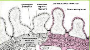
Bee venom phospholipase A2 in the extracellular space near the lipid bilayer. The polar groups of phospholipids are located between the yellow and red planes. Non-polar acyl chains are between the red and black planes.
Pancreatic phospholipase is a digestive enzyme and is involved in the digestion of food lipids. Venom phospholipases are involved in immobilizing the victim due to the lysis of its cells.
2.2 Classification and properties
2.2.1 Cytosolic PLA2
The history of cytosolic phospholipases of group A2 began in 1991, when a protein with a molecular weight of 85 kDa was isolated and cloned from the cytosol of various animal cells, which, in addition to its molecular weight, differed from the phospholipases known at that time in the absence of disulfide bridges and sensitivity to calcium. Later, screening of nucleotide bases made it possible to find two more paralogues – cPLA2b and cPLA2g. The first protein found was named cPLA2a. Called “cytosolic,” the enzyme nevertheless acts on the membranes of the cytoplasmic reticulum and nucleus. This isoform of PLA2 is present in many cells and tissues: brain, kidneys, spleen, lungs, macrophages, neutrophils, alveolar epithelial cells, etc. The enzyme becomes active as a result of phosphorylation by mitogen-activated protein kinases and protein kinase C. Various extracellular cytokines, mitogens, hormones, neurotransmitters, factors growth, antigens, endotoxins, as well as certain physical and stress influences, including ultraviolet light and oxidative stress, induce activation and synthesis of cytosolic PLA2.
The main feature of this enzyme isotype is that it most actively hydrolyzes phospholipid substrates containing arachidonic acid in the second position. This substrate selectivity determines the main function of the enzyme in the cell.
The spatial structure of this enzyme was characterized by nuclear magnetic resonance; X-ray structural data appeared somewhat later. The enzyme hydrolyzes the ester bond at the sn-2 position. To exhibit maximum enzymatic activity, relatively low concentrations of Ca2+ ions are required (about 500 nmol/l); Moreover, the presence of Ca2+ ions is necessary not for the manifestation of enzymatic activity, but for the binding of the protein to the surface of intracellular membranes or, in the case of model systems, to lipid particles. The enzyme practically does not distinguish between groups at the sn-1 position, but has specificity for phospholipids containing arachidonic acid at the sn-2 position. Oleic (18: 1) and linoleic (18: 2) acids are weakly cleaved from the corresponding phospholipids by cPLA2a, but a-linoleic (18: 3) and eicosapentaenoic (20: 5) acids have advantages over arachidonic acid. Since these acids are found in extremely low concentrations in cells, phospholipids with arachidonic acid at the sn-2 position become the main substrate. The enzyme does not show specificity for the substituent at the sn-3 position, but nevertheless diacylglycerol is not its substrate. In addition to the main activity, the enzyme also exhibits lysophospholipase activity, i.e., it is capable of cleaving acyl from the sn-1 position of a lysophospholipid. It is assumed that this activity is necessary to protect the cell from an increased concentration of lysophospholipids, which can disrupt membrane functions. It has been shown that the enzyme can also exhibit transacylase activity activity. The biological significance of this activity is being studied.
Paralogues of cytosolic PLA2 have been described only recently and information about their properties is limited. It is known that the cPLA2g protein does not contain a calcium-binding domain and is independent of Ca2+ ions; has only 29% similarity to the amino acid sequence of the cPLA2a protein. It is membrane bound via a palmitic site and exhibits mainly isophospholipase activity. Its participation in the development of macrophage apoptosis has been shown. The cPLA2b protein has a calcium-binding domain, but also exhibits predominantly the properties of PLA1, i.e. hydrolyzes the sn-1 bond.
The cPLA2a enzyme is currently the only phospholipase that is specific for arachidonic acid and prefers substrates localized in the membrane rather than those in monomeric form in solution. Perhaps these properties can be explained by the existence of an amphiphilic “lid” (amino acid residues 413-457), which prevents the entry of the fatty acid residue of the phospholipid into the active center tunnel if the protein is not localized on membranes. The “lid” opens when the protein interacts with the membrane lipids. The interfacial catalysis carried out by this enzyme has been intensively studied.
Discovery of cytosolic PLA2 in the late 1980s. gave impetus to the study of the regulation of eicosanoid synthesis in the body during an acute response to various proinflammatory stimuli. Regulation of secretory phospholipase occurs at the level of its expression, and the activity of cytosolic phospholipase in the cell is also regulated at the level of enzyme activity. The main factors influencing the activity of cytosolic phospholipase are the concentration of intracellular Ca2+ and phosphorylation of this enzyme by protein kinases. The concentration of Ca2+ ions and the activity of various protein kinases in the cell are very labile parameters that change within a few seconds after the binding of ligands - agonists with the corresponding cellular receptors, which makes it possible to effectively regulate production in the cell arachidonic acid and, accordingly, the synthesis of eicosanoids.
In non-activated cells, the concentration of intracellular Ca2+ usually ranges from 30-100 nmol/l. When various receptors are activated, the concentration of Ca2+ ions can reach 1-3 µmol/l. A number of studies have shown that an increase in the activity of cytosolic PLA2 occurs in the concentration range of Ca2+ ions 150-800 nmol/l. With an increase in the concentration of Ca2+ ions, phospholipase migrates to membranes and attaches to them, after which hydrolysis of phospholipids begins.
The binding of phospholipase to the membrane occurs due to the C2 domain present in the protein:
/>
The C2 domain is homologous to similar domains of protein kinase C, GTPase, and phospholipase C. All these proteins bind to membranes in the presence of Ca2+ ions. The presence of sufficient concentrations of Ca2+ ions is necessary for the orientation of phospholipase and its attachment. In the absence of Ca2+ ions, the enzyme remains active towards soluble substrates, such as 1-palmityl-2-lysophosphatidylcholine.
Depending on the type of agonist, the concentration of Ca2+ ions in the cell changes differently. Some agonists cause its short-term intracellular growth; under the influence of these agonists, the concentration of Ca2+ ions, after a sharp rise, returns to the initial level within 1-2 minutes. Others cause a prolonged increase in the concentration of Ca2+ ions, and the increased concentration of Ca2+ ions remains in the cell for 5-30 minutes. It was shown on epithelial cells that for stable attachment of PLA2 to the surface of biological membranes, an increased concentration of Ca2+ ions is required for 5 minutes, and with a short-term rise after 1-2 minutes, reverse dissociation of the protein into the cytosol occurs without noticeable release of arachidonic acid. If the increased concentration of Ca2+ ions persists for 5 minutes or more, then the cytosolic phospholipase PLA2 remains on the membrane and hydrolysis of phospholipids continues even after the intracellular Ca2+ concentration returns to normal values.
For the manifestation of maximum phospholipase activity with a short-term increase in the concentration of Ca2+ ions inside the cell, phosphorylation of the protein by kinases is also necessary. In vitro experiments have shown that cytosolic phospholipase can be phosphorylated by protein kinase C, p42/p44 mitogen-activated protein kinases, or protein kinase A, but only phosphorylation by mitogen-activated protein kinases (MAPKs) leads to a significant increase in phospholipase activity. Mitogen-activated protein kinases phosphorylate phospholipase at Ser-505. In some cases, it is phosphorylation that determines the manifestation of phospholipase activity in the cell.
Thus, cytosolic phospholipase takes part both in the regulation of eicosanoid synthesis during an acute cell response to various proinflammatory stimuli, and in some cases during a delayed response. The main factors regulating the activity of cytosolic phospholipase are the concentration of intracellular Ca2+ and the activity of mitogen-activated protein kinases. Enzyme activity can increase significantly during the first minutes after cell stimulation. Translocation of the enzyme upon activation is important for two reasons: 1) it allows the enzyme to interact with phospholipids of the membrane; 2) delivers arachidonic acid-selective lipase to the location of the enzymes for the synthesis of prostaglandins (PG) and leukotrienes (LT), i.e., the formation of a compartment is potentially possible in which the released arachidonic acid is directly metabolized further.
2.2.1 Secretory PLA2
Secretory PLA2 have a molecular weight of about 14 kDa, are characterized by the absolute requirement of Ca2+ ions in millimolar concentrations for catalytic activity, the pH optimum is in the range of 7-9.
Currently, ten types of secretory PLA2 have been described (IB, IIA, IIC, IID, IIE, IIF, III, V, X, XII), which differ in the primary structure and location of disulfide bridges. All types of secretory phospholipases are globular proteins rich in cysteine (5-8 disulfide bridges), which ensures the stability of the enzyme, including resistance to proteolysis and denaturation. The enzyme does not show selectivity for the fatty acid composition of phospholipids, but preferentially hydrolyzes negatively charged phospholipids (phosphatidic acid and phosphatidylglycerol).
For a long time, only one PLA2 was known, which is abundantly present in pancreatic fluid (type IB). In 1989, phospholipase type IIA was discovered and cloned, which is stored in secretory granules of platelets and whose concentration increases significantly at sites of inflammation, such as the synovial fluid of rheumatoid arthritis. These proteins are very similar to snake venom proteins. Among the proteins of bee and lizard venoms, other phospholipases were discovered, which are classified as type III. In mammals, a protein corresponding to this type was discovered only in 2000... A new period in research on secretory phospholipases began in 1994, when type IIC and V proteins were discovered. This discovery led to a revision of the role of this family of proteins in the regulation of cell functions and an intensive search for new similar proteins . Proteins of types X, IID, IIE and IIF, XII were discovered.
All of these proteins (except type III protein) have a molecular weight of 14-17 kDa, contain a histidine at the catalytic site, and exhibit phospholipase activity in the presence of millimolar concentrations of calcium. They have highly conserved amino acid sequences in the catalytic region (DXCCXXHD) and the calcium-binding loop region (XCGXGG), as well as 6-8 conserved sulfide bridges. The active center also contains aspartate, which, together with the calcium-binding loop, acts as a pocket for the Ca2+ ion.
Enzymes of this family show no specificity either for the group associated with the phosphatidic acid residue or for the catyl group at the sn-2 position. The presence of oxidized forms of fatty acids in phospholipids increases the activity of secretory phospholipases, which suggests their participation in the regulation of membrane viscosity during oxidative stress. From the X-ray structural analysis of the first two groups of proteins, it follows that there is a hydrophobic channel into which the phospholipid molecule enters after interfacial binding of the protein on the phospholipid surface.
Secretory PLA2 is constitutively contained in various cells involved in the development of immune and inflammatory responses: macrophages, mast cells, fibroblasts and tissues of organs such as the liver, spleen, thymus, bone marrow, and intestines. The activity of the enzyme at the cellular level is regulated due to its induction by various inflammatory stimuli (interleukin-1 and interleukin-6, tumor necrosis factor, lipopolysaccharide, interferon-g, phorbol esters, nerve growth factor). In accordance with induction by various stimulants, the IIA gene promoter contains the nucleotide sequences TATA and CAAT, as well as binding sequences for transcription factors such as AP-1, C/EBP, CREB, NF-kB, STAT, PPA Rg. In some cells, the expression of PLA2 depends on the preliminary activation of the cytosolic phospholipase PLA2a, and the involvement of products of the 12/15-lipoxygenase pathway in the process of regulation of phospholipase IIA is assumed. Glucocorticoids (steroidal anti-inflammatory drugs) are suppressors of phospholipase IIA expression.
Changes in phospholipase IIA expression are in many cases associated with modulation of the prostaglandin branch of the arachidonic acid cascade. Thus, stimulation with interleukin-1 and tumor necrosis factor activated both the synthesis of phospholipase secretion and the synthesis of prostaglandin E2 and prostacyclin in mesangial or endothelial cells, respectively. When antibodies to phospholipase were added to the cells, prostacyclin synthesis was partially suppressed. From the results obtained in recent years, it follows that secretory phospholipases (IIA and similar V, X) are involved in the processes of both rapid and delayed production of arachidonic acid and prostanoids.
It should be noted that the addition of secretory phospholipases to the extracellular environment leads to the active release of arachidonic acid and the synthesis of prostanoids by activated cells, but has virtually no effect on the hydrolysis of membrane phospholipids of resting cells.
In addition to the role of the enzyme responsible for the presence of arachidonic acid, secretory phospholipases can act as physiologically active substances. Thus, in mast cells, specific inhibitors of secretory phospholipases reduced the expression of cyclooxygenase-2 stimulated by nerve growth factor. At the same time, catalytically inactive mutants of the phospholipase protein were also able to induce the expression of cyclooxygenase-2, i.e. this effect does not depend on the enzymatic properties of phospholipase. The mechanism of this process is not clear. The functions of secretory PLA2 as a ligand of specific receptors may be involved.
Indeed, in 1995 it was shown that there are specific proteins that bind to phospholipase type IB (dissociation constant of the resulting complex is 1 nmol/l) and exhibit various biological effects. Over the next 10 years, many more proteins, soluble and membrane-bound, were discovered that are capable of binding secretory phospholipase. However, only one of these proteins exhibits the properties of a “classical” receptor, which upon binding of a ligand activates the intracellular signal transmission system. It is an M-type protein, or sPLA2R. The gene for this protein is localized on the second chromosome; the protein sequence has 75% homology among a number of mammalian species; the gene has 1 copy and is in no way similar to other genes. The protein has a molecular weight of 180-200 kDa, a significant part of it is located in the extracellular region, and a sequence of 40 amino acid residues is located in the cytosol. The protein without this cytosolic region occurs in a soluble form and its role is shown as an inhibitor of the effects of secretory phospholipases. In humans, the protein is expressed in the pancreas, lungs and hips. The important role of activation of this receptor in the development of endotoxic shock has been shown.
The figure shows a diagram of the participation of the secretory phospholipase receptor in the implementation of the biological role of the enzyme at the cellular level.
/>
The diagram shows how active forms of phospholipase sPLA2-IB or sPLA2-X exhibit their enzymatic activity, which leads to the appearance of lipid mediators, and are also high-affinity ligands for the receptor localized in the plasma membrane. The interaction of phospholipase with a membrane receptor leads to the induction of mitogen-activated protein kinases (MAPK) and the corresponding intracellular signal pathway, which stimulates various cell responses: cell proliferation and migration, the synthesis of physiologically active substances. When contact with the membrane is lost, the receptor retains the ability to bind phospholipase, which makes it possible to regulate the activity of the latter as an enzyme and as a ligand.
Thus, secretory phospholipases play a significant role in the development and spread of inflammatory processes in the body. The expression of secretory phospholipases increases significantly in a variety of inflammatory diseases. For this reason, selective inhibitors of the enzymatic activity of proteins of this family are being developed as a potential new class of anti-inflammatory substances. To search for inhibitors of secretory phospholipases, the use of computer modeling methods is promising, since these low-molecular-weight proteins (their molecular weight is about 14 kDa) were obtained in a crystalline state, and their three-dimensional structures are known. There is reason to believe that in the next 5-10 years, based on the results of these studies, new therapeutic technologies and new drugs will be created.
2.2.3 Calcium-independent PLA2
Classic calcium-independent PLA2 (iPLA2-VIA) exists in a voligomeric form and has several splice variants, at least two of which (VIA-1 and VIA-2) have enzymatic activity. The iPLA2-VIA-1 protein has a molecular weight of 85 kDa; contains 8 ankyrin repeats in the N-terminal region, a catalytic domain with a characteristic amino acid sequence GXSXG, where S - serine-465 acts as the center of catalysis. There is also an ATP-binding sequence GXGXXG. Near the C-terminus (in the region of amino acid residues 694-705) there is a binding region for calmodulin. The formation of a complex of the iPLA2-VIA protein with activated calmodulin (i.e., in the presence of Ca2+ ions) leads to the inactivation of this phospholipase. Although both splice forms VIA-1 and VIA-2 have ATP-binding sequences, only iPLA2-VIA-2, but not iPLA2-VIA-1, has been shown to be activated severalfold in the presence of ATP.
Ankyrin repeats are found in several hundred proteins, such as transcription factors and cell cycle regulators. This motif is believed to be involved in protein-protein interactions. It is assumed that iPLA2-VIA protein is present in cells as a tetramer and, possibly, ankyrin motifs are involved in protein oligomerization. Phospholipase iPLA2-VIA is not specific to the nature of the fatty acid at the sn-2 position and the substituent at the sn-3 position of the phospholipid; It is fully active in the absence of calcium and performs phase-transfer catalysis. The enzyme also exhibits lysophospholipase activity at the sn-1 position, transacylase activity, and activity characteristic of the PAF-AH protein.
The iPLA2-VIB protein contains a GVSTG lipase motif, where serine-483 is located in the catalytic center; ATP-binding motif; signal localization motif in peroxisomes at the C-terminus of the molecule. The C-termini of the molecules, including the ATP-binding repeat and the catalytic region, of the iPLA2-VIB and iPLA2-VIA proteins are similar. Differences are observed in the N-terminus of the protein, where the iPLA2-VIB protein has non-tankyrine motifs, many serine and threonine residues, some of which can be phosphorylated by protein kinases A and C. There are also five Ser-Pro regions that are targets for proline kinases. One of these sequences (PTSP, residues 269-272) is a site of phosphorylation by mitogen-activated protein kinases. The activity of this isoform does not depend on calcium ions. The presence of different phosphorylation sites in proteins of the iPLA2-VI group indicates that the roles of the iPLA2-VIA and iPLA2-VIB proteins in cells may differ. Both calcium-independent phospholipases (iPLA2-VIA and iPLA2-VIB) are membrane-bound proteins.
2.3 Substrate specificity
Catalysis by the action of PLA2 is characterized by the surface, positional and steric specificity of the enzyme. Phospholipase catalyzes the reaction at the lipid/water interface. On the one hand, for most lipolytic enzymes, including PLA2, the enzyme activity is significantly higher when substrates exist in the form of aggregates (micelles, mixed micelles, monolayers and bilayers) than when acting on water-soluble substrates in monomolecular form.
On the other hand, PLA2 does not act on lipid molecules in densely packed and, therefore, hard-to-reach lipid aggregates. To create a phase interface in these cases, detergents (Triton x-100, sodium deoxycholate) are usually used. It has been proven that for optimal manifestation of the surface specificity of the enzyme, the presence of aggregated substrates with a certain acyl chain length is necessary.
With the discovery of the steric and positional specificity of PLA2, rather strict minimum requirements of the enzyme for the substrate were formulated: in the sn-2 position of the glycerol residue, the lipid must contain an ester group, and in the sn-3 position - a phosphate group. Later it was shown that the phosphoric acid residue can be replaced by a sulfonium acid: sulfolipid turned out to be a good substrate of PLA2, the corresponding phospholipids, where the C-O-P bond is replaced by a C-P bond, are also hydrolyzed by the enzyme, but to a lesser extent. Therefore, the minimum requirements of the enzyme for the substrate are currently reduced to the fact that the glycerophospholipid has a natural L-configuration and has an ester bond in the sn-2 position of glycerol, and also contains a group with strong anionic properties at a distance of five to six atoms from the carboxyl carbon. It is believed that the phosphate group must have one free acid function.
In a phospholipid with two sensitive ester bonds - diphosphatidylglycerol (cardiolipins) - both can be hydrolyzed. It was found that β-lecithins (1, 3-diacyl-sn-glptsero-2-phosphocholine) are hydrolyzed by PLA2 from the snake snake Crotalusadaincniteus, although at a relatively low rate. PLA2 does not hydrolyze the amide bond of sphingolipids. Thiol analogues of phosphatidylcholines are good substrates, which makes it possible to monitor the hydrolysis process spectrophotometrically. De Haas et al. found that a negative charge on the phosphate group is not an absolute requirement for the substrate.
Due to its steric and positional specificity, PLA2 is a valuable tool in lipid chemistry and biochemistry. They are used to establish the positional distribution of fatty acids in the analysis of phosphoglycerides, to separate racemic mixtures of lipids, as well as in lipid synthesis to obtain phosphoglycerides with a mixed composition of fatty acids.
The substrate specificity of cellular PLA2 has not yet been fully delineated, however, data on the structure of substrates for PLA2 cells in the blood and immune system have been accumulated; in particular, the specificity of these enzymes for phosphatidylcholines containing arachidonate in the sn-2 position of glycerol has been proven.
2.4 PLA2 inhibitors
2.4.1 Non-competitive inhibitors
A decrease in the catalytic activity of PLA2 can occur due to an imbalance at one or several stages of the enzymatic reaction, therefore, known inhibitors of this enzyme can be roughly divided as follows.
a) Binding by an enzyme inhibitor (E) can shift the E↔E* equilibrium to the left and reduce the concentration of catalytically active E*. This occurs when the substrate vesicles are supplemented with vesicles of a non-hydrolyzable analogue of the substrate, with which the enzyme binds but cannot be rapidly desorbed. The need for the presence of calcium ions during the binding of PLA2 to the interfacial surface gives grounds to classify chelating agents, such as EDTA, as inhibitors.
b) Lipophilic compounds change the phase properties of substrates and reduce the charge density on the interphase surface, shifting the E↔E* equilibrium to the left. It has been shown that such changes are caused by organic solvents, detergents, alcohols, as well as cationic amphiphiles, phenothiazines and local anesthetics of various chemical natures. Other inhibitors - fatty acids, mepacrine, aristolochic acid - also affect the binding-desorption stage of E↔E* without affecting the catalytic effect of the enzyme on the interfacial surface.
c) Some types of nonspecific inhibition. Under certain conditions, the rate of hydrolysis under the influence of PLA2 of phospholipids that have undergone peroxidation increases significantly. Therefore, antioxidants are considered to have the potential to reduce enzyme activity. Proteins such as lipocortin and calpactin solubilize phospholipids of the interfacial surface, thereby reducing PLA2 activity. Water-soluble anions, such as heparin, inhibit the binding of PLA2 to the interface by blocking the anionic binding site of the enzyme.
d) Covalent modifications of amino acid residues of PLA2. 1-Bromoctan-2-one and n-bromophenacyl bromide covalently bind to His-48 in the catalytic center of the enzyme and completely inhibit catalytic activity; the rate of such modification is significantly reduced when the enzyme is already bound to the interfacial surface. Dialdehydes like gossypol modify the amino groups of PLA2 lysine residues responsible for its interfacial binding; the rate of modification increases in the case of an enzyme already associated with the interfacial surface. The non-steroidal terpenoid manoalide, isolated from a sea sponge, and its synthetic analogue manoalogue act in a similar way.
e) Other connections. Presumably, many other compounds, including some drugs, inhibit PLA2 activity in vivo, however, the mechanism of their inhibitory action has not yet been elucidated. Among these substances are bioflavonoids and retinoids, hydroxyeicosatetraenoic acids, phenofetol (β-adrenergic receptor agonist), gabexate mesylate, nisergoline, papaverine, cinnarizine and amperone.
2.4.2 Competitive inhibitors
Competitive inhibitors of PLA2 are analogues of substrates, reaction products or transition state complexes. They compete with the substrate for binding to the active center of the E* molecule, effectively reducing the concentration of the ES* complex. This mechanism of inhibition was experimentally confirmed when studying the catalysis of phospholipids under the influence of PLA2 according to the “scooting” type.
A common strategy for creating inhibitors is to replace the PLA2-sensitive ester linkage with a non-hydrolyzable group. In this case, the inhibitor must remain a close structural analogue of the substrate and not cause changes in lipid membranes. At the moment, it is not possible to compare the available data on competitive inhibitors, since a unified theory and quantitative description of lipolysis at the lipid/water interface has not yet been developed.
Aminoacyl phospholipids. Among phosphatidylcholines with modification of the sn-2-ester linkage, a class of powerful competitive inhibitors of PLA2 - sn-2-amide analogues - was discovered. Replacing the sn -2-ester bond with an ether bond or with a hydrocarbon residue also resulted in competitive inhibition of PLA2, but to a lesser extent.
The effect of aminoacyl analogues of phospholipids on the enzymatic activity of PLA2 was studied mainly in model experiments with mixed micelles under the following conditions:
1) the total concentration of lipids (inhibitor and substrate) [I] + [S] must be constant to calculate the mole fraction of the inhibitor, α = [I]/([I] + [S]);
2) the substrate and inhibitor molecules must occupy the same area on the interfacial surface;
3) the interfacial surface of the micelles must be large enough so that the enzyme is only in a bound form.
To assess the effect of the inhibitor on the activity of PLA2, the conditional value of the inhibitory strength (Z) was used, which is a measure of the ratio of the interfacial dissociation constants for the substrate and the inhibitor and is determined by the expression:
Rv= I + αZ,
where Rv is the ratio of the reaction rate at K≠Km to the reaction rate at Ki=Km, α is the mole fraction of the inhibitor in the micelle.
The inhibitory effect of (R)-1-alkyl-2-acylamino-1,2-dideoxyglycero-3-phosphocholine analogues on mammalian pancreatic phospholipases was also studied. Among inhibitors saturated with fatty acid chains, analogues with C10 acyl chains had the greatest activity. The behavior of unsaturated analogues was more complex in both zwitterionic and anionic inhibitors; an increase in the number of cis-double bonds at their particular location on the vacyl chain led to an increase in the Z parameter.
In the course of studies with thioamide analogues of substrates, it turned out that the thioamide analogue of phosphatidylethanolamine with lC50 = 4.5 * 10-7 M is the most powerful known PLA2 inhibitor.
Studies conducted with aminoacyl inhibitors have revealed several aspects of the enzyme/lipid interaction:
1) The introduction of an amide residue into the sn-2 position of a phospholipid significantly increases its binding to the catalytic center of PLA2: the more nucleophilic oxygen of the amide group is able to interact more strongly with the electrophile of this center (presumably Ca2+). The amide group also provides better opportunities for hydrogen bonding.
2) The α-methylene group of the acyl residue at the sn-2 position of the phospholipid is responsible for the binding of the phospholipid to the catalytic center of the enzyme.
3) An increase in the hydrophobicity of the functional group at the sn-1 position of the phospholipid increases the affinity between the enzyme and the substrate.
4) Phosphatidylethanolamines turned out to be more powerful inhibitors than phosphatidylcholines.
The approach to the synthesis of optically active 1-acyl-2-acylamino-2-deoxyglycerophosphocholines is based on maintaining the chirality of the starting compound (L-serine) throughout the synthesis. The selected sequence of introduction of substituents involves the use of a minimal number of protecting groups. A stereospecific method for the synthesis of 1-alkyl-2-acylamino-2-deoxyglycerophosphocholines from L-serine has also been developed. The introduction of an aliphatic alkyl group is carried out by the interaction of a fatty acid methanesulfonate with an oxazoline-protected deoxyglyceride. Racemic long-chain acylamino analogues of phospholipids were proposed to be obtained starting from 2-aminopropanol. The synthesis of an optically active thioamide analogue of phosphatidylcholine is described.
Phtarketone analogues. It has previously been established that substrate analogues containing polarized ketone groups, including fluoroketone and 1,2-diketone groups, inhibit hydrolytic enzymes. The best inhibitor was a substituted phosphoethanolamine with a single acyl residue, despite the fact that the enzyme prefers substrates with two acyl residues
Since the difluoroketone group is easily hydrated in an aqueous solution, the inhibitors resemble in structure the tetrahedral intermediate that is formed during lipolysis.
Among the fluoroketone analogues, selective inhibitors of intracellular cytosolic PLA2 have been found. These inhibitors turned out to be electrophilic ketone analogues of arachidonic acid.
The most powerful inhibitor was α-trifluoromethyl ketone of arachidonic acid. The structure of the complex of this substance with PLA2 was analyzed using 19F and 13C NMR spectroscopy. The results confirmed the hypothesis that binding to the active center of the enzyme forms a hemiketal with a serine or threonine residue of the protein molecule, due to the ability of α-fluoroketones to easily hydrate in aqueous solutions.
Phosphate analogues. To study the nature of PLA2 inhibition from various sources and the effect of substituents on the inhibitory ability, more than 100 sn-2-phosphate analogues of phosphatidylcholines were synthesized. Compounds of this class inhibited only the enzyme already associated with the interfacial surface and had no effect on the desorption of the enzyme. Phosphate inhibitors bind to the active center of the enzyme through the calcium ion through the coordination bond E-Ca... O=P, competing with substrates. This interaction is modulated by substituents on the inhibitor molecule. The replacement of the O atom in this complex with S, NH2, the 0=P group with O=C-O, and the presence of a negatively charged phosphate group significantly reduced the affinity for the enzyme. The inhibitory ability of phosphoesters was strictly dependent on stereochemical and structural features: the chirality of the sn-2 position, the length of the alkyl chain in the sn-position, and the presence of a hydrophobic substituent in the sn-3 position of glycerol. Sulfonate, amide, oxime-containing, dianionic phosphomonoester analogues did not exhibit inhibitory properties.
Alkyl phosphotidylcholines. For pharmacological use, apparently, the most promising substances are those that can inhibit PLA2 without disrupting the structural organization of the membrane. Of particular interest in this regard are lipids with ether bonds. In lipid molecules of this class, hydrophobic substituents are attached to the hydrophilic residue through a non-hydrolyzable PLA2 ether bond. At the same time, in their composition, membranelipids with an ether linkage are practically no different from their diacyl analogues. We studied the effect of long-chain ether-linked phosphatidylcholines on the destruction of membranes under the influence of PLA2 using P-NMR spectroscopy and assessed the possibility of using lipids in local inflammatory reactions.
Almost identical results were obtained for the studied inhibitors: the introduction of compounds into the lipid bilayer from egg phosphatidylcholine in a molar ratio of 1: 1 led to such effective membrane stabilization that no structural rearrangements were observed under the influence of this enzyme.
Among the phospholipids of this class, inhibitors of cytosolic PLA2 were found, which have anti-inflammatory and antiallergic properties.
2.5 Importance for the body in case of impaired activity
Excessive activation of PLA2 is important in the pathogenesis of cell damage. Unsaturated fatty acids (arachidonic, pentanoic, etc.) released under the action of phospholipase are consumed to form physiologically active compounds - prostaglandins and leukotrienes. The remaining part of the phospholipid molecule (lysophospholipid) has only one fatty acid “tail”, as a result of which it has the ability to form micelles and is a very strong detergent. Damage to cell membranes under conditions of excessive activation of PLA2 is associated with the detergent effect of lysophospholipids.
Pharmacological and toxic properties of phospholipase due to its biochemical activity. With the help of its toxic lysolecithin, it breaks down blood and tissue structures, damaging their cell membranes and organelles. Phospholipase reduces blood clotting by destroying clotting-promoting components that contain phospholipids. It damages mitochondrial membranes, and the latter are an important cell organelle, carriers of complex enzyme systems. They participate in metabolism and redox processes. Damage to the phospholipid structure of nerve fibers is likely caused by phospholipase, which blocks conduction between nerve and muscle tissue.
The introduction of phospholipase under the skin causes local inflammation; intravenous administration is accompanied by a decrease in blood pressure in experimental animals, pulmonary edema, and hemorrhages in the alveoli. In animals that have survived for several hours, hemoglobin from destroyed red blood cells is detected in the urine. In addition, it was found that bee venom phospholipase, injected subcutaneously, enhances the development of model inflammatory processes of various etiologies.
It has been suggested that, through its surface tension-lowering effect, melittin prepares phospholipids for the enzymatic activity of phospholipase. Of all the components of bee venom, phospholipase is the most powerful antigenic and allergenic irritant. The blood of beekeepers immune to bee venom contains antibodies with a high titer against the venom. Patients hypersensitive to bee venom showed an acute reaction to phospholipase during laboratory tests for allergy.
The toxic and allergic properties of phospholipase that enhance inflammatory processes characterize it as a component of bee venom that is harmful to the human body.
Activation of PLA2 is calcium dependent. It occurs when adrenal cells are stimulated, which leads to an acceleration of arachidonylphosphatidylinositol turnover. This effect is also caused by a calcium ionophore and may reflect an increase in intracellular calcium levels and secondary stimulation of PLA2 as an early reaction accompanying receptor interaction. It is known that the effect of nasteroidogenesis in the adrenal glands depends on calcium, and not only on the formation of cAMP. At least part of the calcium requirement may be related to PLA2-co-mediated turnover of membrane phospholipids during activation of the adrenal cortex.
The mechanism, including activation of phospholipase, may reflect a general property of hormone-regulated secretory cells; with hormonal stimulation of specific target cells, other stages of phospholipid metabolism also change. Thus, in ovarian granuloma cells, where LH increases the production of prostaglandins, the hormone does not increase the formation of arachidonic acid, but acts at later stages , increasing the activity of prostaglandin synthetase. This effect of LH on prostaglandin synthesis in the Graafian follicle (vesicular ovarian follicle) does not appear to mediate the steroidogenic effects of gonadotropin, but plays an important role in the development of ovulation.
2.6 Use of PLA2 in medicine
Secretory PLA2 is considered as one of the pathogenetic factors in the formation of a number of diseases: rheumatoid arthritis, atherosclerosis. In recent years, information has emerged about the involvement of this enzyme in lung pathology. Of particular interest is the pathogenetic chain “sPLA2 – acute respiratory distress syndrome (ARDS) – lung surfactant.”
Acute respiratory distress syndrome in adults occurs as a result of both direct effects on the lungs (aspiration, inhalation of toxic substances, 100% oxygen, insufficiently qualified mechanical ventilation) and indirect effects (sepsis, shock of any etiology, polytrauma, blood loss). Despite years of intensive research, the mechanism of development of ARDS remains unclear, and its mortality rate is high (~50%). It is believed that the origin of this lung condition is a decrease in the production and activity of surfactant.
Lung surfactant is a lipoglycoprotein complex that is synthesized by type II alveolocytes. It consists of 80-90% phospholipids, 5-10% neutral lipids and 5-10% proteins. In addition to surface-active properties necessary for normal breathing, it has anti-inflammatory and immunoregulatory effects. Violation of its integrity leads to an increase in surface tension not only in the alveoli, but also in the bronchioles and small bronchi.
The substrate specificity of cytosolic PLA2 to arachidonoyl-containing substrates suggests that this isoform plays a major role in the pathogenesis of asthma. It is known that leukotrienes B4, C4, D4, E4 are the main group of mediators involved in the complex inflammatory process and leading to clinical manifestations of asthma. The action of leukotrienes is to contract bronchial smooth muscle, increase the amount of mucus produced, stimulate vascular permeability and the formation of edema. All these signs are characteristic of asthma. Leukotrienes are synthesized in response to exposure to an allergen or a nonspecific reaction leading to activation of cPLA2.
Chilton F.H. studied the presence of phospholipase activity products in the bronchoalveolar lavage fluid of patients with asthma 5-30 minutes, 6, 20 hours after antigen challenge. The concentration of lysoproducts was 7 times higher compared to the control group. Hence, it was suggested that the hydrolysis of surfactant phospholipids leads to the generation of cytotoxic lysophosphatidylcholine, which can have a direct detergent effect on the membrane and influence the activity of membrane channels and proteins. In addition, transforming into platelet activating factor, it causes disruption of the permeability of the alveolar-capillary barrier, bronchospasm, and platelet aggregation.
It is also assumed that cPLA2 is involved in the occurrence and development of pulmonary fibrosis, a currently poorly understood interstitial lesion of the lung parenchyma. The pathogenesis of this condition includes excessive production of proinflammatory mediators, alveolar inflammation, fibroblast proliferation, and collagen accumulation. The classic experimental model for this condition is pulmonary fibrosis induced by intratracheal or intraperitoneal administration of bleomycin.
The role of phospholipase in the development of some forms of pancreatitis is great.
With insufficiency of the major duodenal papilla and increased pressure in the duodenum, reflux of bile or duodenal contents into the pancreatic ducts is possible. Bile, entering the pancreatic ducts, can be exposed to pancreatic phospholipase, already activated by trypsin, resulting in the formation of lysolecithin, which, penetrating into the interstitial space of the pancreas, causes deep damage in the cells.
Also, Dr. Heidi May proved that lipoprotein-associated PLA2 predicts the risk of coronary death.
Information obtained in recent years about the involvement of cytosolic and secretory PLA2 in the formation of lung pathology opens up the prospect of new approaches to the treatment of such diseases. However, first it is necessary to clearly define the place of these enzymes in the pathogenetic chain of molecular events. Here the “syndromic” approach may be important. For example, there is still no information about the state of PLA2 during hyperthermia and hypoxia, or during activation of producer cells. Meanwhile, most lung diseases are characterized by these conditions, which largely determine the clinical picture, course and outcome of the pathological process.
2.7 Biological role of PLA2
To date, the secreted PLA2 of mammalian venoms and pancreatic glands has been sufficiently fully characterized and studied. In contrast, the relatively low in vivo concentrations of intracellular and nonpancreatic extracellular PLA2 seriously complicate studies of this class of enzymes.
It has been established that membrane-bound PLA2 plays an important role in the regulatory processes of cellular metabolism. Several pathways for the regulation of these enzymes are known, but the general mechanism is very complex and not fully understood. PLA2 is involved in the transmission of chemical signals through membranes in response to external influences. The enzyme effectively hydrolyzes phospholipids containing fatty acid peroxides, restoring the structural and functional properties of cell membranes. Apparently, a change in the molecular conformation of oxidized lipids facilitates the enzyme's access to the sn-2-ester bond.
Today it has been established that phospholipases A2 play a significant role in the development of the inflammatory process. The contribution of the enzyme is to trigger the synthesis of lipid regulators of this reaction - one of the groups of so-called chemical mediators of inflammation. They are formed, activated or mobilized in the inflammatory focus and their relationship determines the nature of the pathological process. Lipid inflammatory mediators are represented by fatty acids and their derivatives (prostaglandins, leukotrienes, thromboxanes), as well as phospholipid platelet activating factor (PAF). It is believed that intracellular cytosolic PLA2 is involved in the inflammatory process, releasing polyenoic acids from the sn-2 position of the glycerol residue of membrane phospholipids. Polyenoic fatty acids, including arachidonic acid, have their own biological activity, incl. increase vascular permeability, cause platelet aggregation, and have a vasoactive effect.
Other products of the phospholipase hydrolysis reaction are lysophospholipids, which have pronounced cytotoxicity and detergent properties. These compounds are found in diseases such as cholecystitis, myocardial infarction, cataracts, psoriasis, etc. When a fatty acid is released from 1-O-alkyl-type phosphatidylcholine, the resulting lysophospholipid serves as a precursor to PAF, a mediator of inflammation, allergic reactions, septic shock and asthmatic conditions. This compound is currently being intensively studied; a large number of publications have been devoted to PAT agonists and antagonists.
The importance of membrane-bound PLA2 in cellular regulation, as well as its elevated level in a number of pathological processes, lead to the need to regulate the activity of this enzyme. Therefore, the search for new classes of lipid inhibitors and the development of convenient methods for their synthesis are currently of practical interest.
Bibliography
1. Bragina N.A., Chupin V.V., Bulgakov V.G. Lipid inhibitors of phospholipase A2, M., 1999, p. 83-96
2. Brockerhoff X., Jensen R., Lipolytic enzymes, trans. from English, M., 1978, p. 242-356
Phospholipase- an enzyme that hydrolyzes phospholipids. There are 4 main classes of phospholipases: A, B, C and D, each of which hydrolyzes a specific bond in the phospholipid. Class A phospholipases are divided into phospholipases A1, which cleave off the SN-1 acyl chain of a phospholipid, and phospholipases A2, which cleave off the SN-2 acyl chain. Phospholipases A2 include:
- extracellular phospholipases A2 of snake, reptile and insect venoms
- pancreatic phospholipase
- intracellular phospholipases A2
Pancreatic phospholipase
Pancreatic phospholipase- class A2 phospholipase (EC 3.1.1.4), a lipolytic enzyme that decomposes food fats (triglycerides). It is secreted in the form of the pancreatic proenzyme prophospholipase, which is activated in the small intestine by trypsin (Sablin O.A. et al.). The optimal acidity for the catalytic activity of pancreatic phospholipase is 7.5–9.0 pH. “Works” as an enzyme in the presence of Ca 2+ (which is a coenzyme for phospholipase).When bile lecithin is hydrolytically broken down by phospholipase A, lysolecithin is formed. Further metabolism of lysolecithin is catalyzed by phospholipase B, which is inhibited by bile acids (Maev I.V. et al.)
The role of phospholipase in the development of some forms of pancreatitis
With insufficiency of the major duodenal papilla and increased pressure in the duodenum, reflux of bile or duodenal contents into the pancreatic ducts is possible. Bile, entering the pancreatic ducts, can be exposed to pancreatic phospholipase, already activated by trypsin, resulting in the formation of lysolecithin, which, penetrating into the interstitial space of the pancreas, causes deep damage to the cells.Table of contents of the topic "Endocrine system. Chemical nature and general mechanisms of action of hormones.":
1. Endocrine system. Chemical nature and general mechanisms of action of hormones.
2. Mechanisms of action of peptide, protein hormones and catecholamines. Ligand.
3. Basic systems of secondary intermediaries. Adenylate cyclase system - cAMP.
5. Calcium-calmodulin system. Interconnections of secondary intermediaries.
6. The mechanism of action of steroid hormones. Genomic mechanism of action.
7. Non-genomic mechanism of action of steroid hormones.
8. Self-regulation of effector sensitivity to hormonal signals. Desensitization (reduced sensitivity) of the cell.
9. Regulatory functions of pituitary hormones. Adenohypophysis. Neurohypophysis.
10. Blood supply to the adenohypophysis and neurohypophysis.
Guanylate cyclase-cGMP system. Activation of membrane guanylate cyclase does not occur under the direct influence, but indirectly through ionized calcium and oxidant membrane systems. The stimulation of guanylate cyclase activity, which determines the effects of acetylcholine, also occurs indirectly through Ca2+. Through activation of guanylate cyclase, the effect of atrial natriuretic hormone, atriopeptide, is realized. By activating peroxide oxidation, the vascular wall endothelial hormone nitric oxide, a relaxing endothelial factor, stimulates guanylate cyclase. Under the influence of guanylate cyclase from GTP is synthesized by cGMP, activating cGMP-dependent protein kinases, which reduce the rate of phosphorylation of myosin light chains in the smooth muscles of the vascular walls, leading to their relaxation. In most tissues, the biochemical and physiological effects of cAMP and cGMP are opposite. Examples include stimulation of cardiac contractions under the influence of cAMP and inhibition of contractions by cGMP, stimulation of contraction of intestinal smooth muscles by cGMP and inhibition of cAMP. cGMP ensures hyperpolarization of retinal receptors under the influence of light photons. Enzymatic hydrolysis of cGMP, and consequently the cessation of the hormonal effect, is carried out using a specific phosphodiesterase.
Rice. 6.2. Mediation of hormonal signals by the phospholipase C-inositol-3-phosphate system.The formation of a hormone-receptor complex with the participation of a regulatory G protein activates membrane phospholipase C, which causes the hydrolysis of membrane phospholipids with the formation of two secondary messengers: inositol-3-phosphate and diacylglycerol. Inositol-3-phosphate leads to the release of Ca2+ from intracellular stores. The binding of ionized calcium to the specialized protein calmodulin activates protein kinases and causes phosphorylation of intracellular structural proteins and enzymes. Diacylglycerol increases the affinity of protein kinase C for Ca2+, promoting its activation, which also results in protein phosphorylation. Diacylglycerol simultaneously implements another way of mediating the hormonal effect, activating phospholipase A-2 and the formation of prostanoids.
Phospholipase C system - inositol-3-phosphate.
Hormone receptor complex with the participation of the regulatory G protein leads to activation of the membrane phospholipase C enzyme, causing hydrolysis of membrane phospholipids with the formation of two secondary messengers: inositol 3-phosphate and diacylglycerol.Inositol 3-phosphate causes the release of Ca2+ from intracellular stores, mainly from the endoplasmic reticulum, ionized calcium binds to the specialized protein calmodulin, which ensures the activation of protein kinases and phosphorylation of intracellular structural proteins and enzymes. In turn, diacylglycerol promotes a sharp increase in the affinity of protein kinase C for ionized calcium, the latter activates it without the participation of calmodulin, which also ends with the processes of protein phosphorylation. Diacylglycerol simultaneously implements another way of mediating the hormonal effect by activating phospholipase A-2. Under the influence of the latter, arachidonic acid is formed from membrane phospholipids, which is a source of substances with powerful metabolic and physiological effects - prostaglandins and leukotrienes. In different cells of the body, one or another pathway for the formation of secondary messengers prevails, which ultimately determines the physiological effect of the hormone. Through the considered system of second messengers, the effects of adrenaline (in connection with the alpha-adrenoreceptor), vasopressin (in connection with the V-1 receptor), angiotensin-I, somatostatin, and oxytocin are realized.
Page 1
Phospholipase A2 (E.F.3.1.1.4.) is an enzyme that catalyzes the cleavage of fatty acid residues - lecithin, cephalin - from phospholipids, converting them into toxic compounds that greatly reduce surface tension. These compounds dissolve red blood cells and other cellular and subcellular structures and are therefore called lysolecithins and lysokephalins
In the phospholipid molecule, bee venom phospholipase cleaves off the fatty acid from the second place in the molecule and is therefore called phospholipase A2. It has been known since 1897 (Langer) as a factor that enhances the hemolytic activity of bee venom after the addition of lecithin. Phospholipase is the most studied enzyme in bee venom. Phospholipase was discovered as a digestive enzyme in 1900. It is now clear that phospholipase A2 (PLA2) is more than a digestive enzyme. It is widespread and present in most mammalian cells and tissues, serving as a regulator of metabolism, maintaining membrane homeostasis, and the formation of eicosanoid precursors.
Depending on the molecular weight, cellular localization and the presence of Ca2+ ions, cytosolic PLA2, secretory and Ca-independent PLA2 or outer membrane PLA2 are distinguished.
PLA2s comprise several unrelated protein families with common enzymatic activities. The two most important families are secreted and cytosolic phospholipases A2.
Cytosolic PLA2:
Intracellular phospholipases, as well as extracellular ones, are calcium-dependent enzymes. Structurally, however, they are very different from secreted phospholipases. Typically, they are much larger (more than 700 amino acids) and contain a C2 domain, which directs the enzyme to the cell membrane. These phospholipases are mainly involved in cellular signaling pathways such as the inflammatory response. Under the influence of PLA2, arachidonic acid, a precursor of eicosanoids, active signaling molecules such as leukotrienes and prostaglandins, can be formed in the cell.
PLA2 outer membrane:
Gram-negative bacteria contain PLA2 on their outer membrane with a wide range of specificity. In Escherichia coli, this enzyme is involved in the release of the bacteriocin toxin from the cell due to increased membrane permeability with an increase in the level of lysophospholipids and fatty acids in the membrane.
Secreted PLA2:
Extracellular forms of phospholipases have been isolated from various venoms of snakes, bees and wasps. They are also found in all mammalian tissues and in bacteria. The activity of these phospholipases requires the presence of calcium.
Bee venom phospholipase A2 in the extracellular space near the lipid bilayer. The polar groups of phospholipids are located between the yellow and red planes. Non-polar acyl chains are between the red and black planes.
Pancreatic phospholipase is a digestive enzyme and is involved in the digestion of food lipids. Venom phospholipases are involved in immobilizing the victim due to the lysis of its cells.
The history of cytosolic phospholipases of group A2 began in 1991, when a protein with a molecular weight of 85 kDa was isolated and cloned from the cytosol of various animal cells, which, in addition to its molecular weight, differed from the phospholipases known at that time in the absence of disulfide bridges and sensitivity to calcium. Later, screening of nucleotide bases made it possible to find two more paralogs – cPLA2b and cPLA2g. The first protein found was named cPLA2a. Called “cytosolic,” the enzyme nevertheless acts on the membranes of the cytoplasmic reticulum and nucleus. This isoform of PLA2 is present in many cells and tissues: brain, kidneys, spleen, lungs, macrophages, neutrophils, alveolar epithelial cells, etc. The enzyme becomes active as a result of phosphorylation by mitogen-activated protein kinases and protein kinase C. Various extracellular cytokines, mitogens, hormones, neurotransmitters, growth factors, antigens, endotoxins, as well as certain physical and stressful influences, including ultraviolet light and oxidative stress, induce activation and synthesis of cytosolic PLA2.
Introduction
1. Phospholipases
1.1 Classification. Properties
2. Phospholipase A2
2.1 General information (reaction, discovery, structure)
2.2 Classification and properties
2.2.1 Cytosolic PLA2
2.2.2 Secretory PLA2
2.2.3 Calcium-independent PLA2
2.3 Substrate specificity
2.4 PLA2 inhibitors
2.4.1 Non-competitive inhibition
2.4.2 Competitive inhibition
2.5 Implications for the body in case of activity impairment
2.6 Use of PLA2 in medicine
2.7 Biological role of PLA2
Bibliography
Introduction
Phospholipases are enzymes of the class of hydrolases that catalyze the hydrolysis of phosphoglycerides. Depending on the position of the hydrolyzed bond in the phospholipid, there are 4 main classes of phospholipases: A, B, C and D.
Lysophospholipids are cleaved by phospholipases L (the existence of position-specific phospholipases L1 and L2 has not been proven). Phospholipase B is an obsolete name. drugs with phospholipase A and L type activity.

X - residue of choline, serine, myoinositol, etc.; for phospholipases L1 R2=C(O)R4, R3=H; for phospholipases L2 R2=H, R3=C(O)R4
Each of the phospholipase families is heterogeneous and includes enzymes that differ significantly in molecular weights, subunit composition and other properties. All phospholipases most actively catalyze hydrolysis at the phospholipid-water interface; slowly hydrolyze water-soluble substrates.
Phospholipase A1- (EC 3.1.1.32, English phospholipase A1) cleaves off the SN-1 acyl chain.
Phospholipase A2- (EC 3.1.1.4, English phospholipase A2) cleaves off the SN-2 acyl chain.
Phospholipase B-(lysophospholipase, English phospholipase B) cleaves off both SN-1 and SN-2 acyl chains. Phospholipase, which has the activities of both phospholipase A1 and A2, that is, capable of hydrolyzing the acyl chain of a phospholipid in the sn-1 and sn-2 positions.
Phospholipase C- (EC 3.1.4.3, English phospholipase C) hydrolyzes the bond between the glycerol residue of the phospholipid and the polar phosphate group, resulting in the formation of diacylglycerol and a phosphate-containing polar group.
Phospholipase D- (EC 3.1.4.4, English phospholipase D) hydrolyzes the bond between the phosphate group and the alcohol group, releasing phosphatidic acid and alcohol. There are 2 isoforms of this phospholipase D1 and D2.
Phospholipases play an important role in lipid metabolism in living organisms. They are used to determine the structure of phosphoglycerides and their location in membranes.
1. Phospholipases.
1.1 Classification. Properties
In fact, several group A phospholipases are distinguished; they are an integral part of many tissues and secretions of living organisms.
Phospholipase A1 For the most part, intracellular enzymes, often membrane-bound, do not require coenzyme. Their molecular weights vary between 15-90 thousand; optimal catalytic activity occurs at pH 4.0 (for lysosomal enzymes) or 8.0-9.5 (for enzymes of microsomes, plasma membranes and cytosol); widely distributed in animal tissues (liver, heart, brain) and in microorganisms (Bacillus subtilis, B. megateiium, Mycobacter phlei, Escherichia coli).
Phospholipases A1 cleave off the phospholipid acyl chain at the sn-1 position. The action of phospholipase A1 on a phospholipid produces 1-lysophospholipid and a fatty acid. Phospholipase is an active component of snake venom with hemolytic action.
Phospholipase A2- the most studied representatives of phospholipases. There are 3 known groups of phospholipases A2: 1) enzymes from the venoms of snakes, reptiles and insects, existing in the form of a large number of isoforms; 2) enzymes of the mammalian pancreas, produced in the body in the form of zymogens (precursors with a higher molecular weight) and activated by trypsin; 3) intracellular enzymes from the blood and tissues of animals, among which there are both soluble and membrane-bound.
Phospholipases A2 of the first two subgroups are water-soluble enzymes with a molecular weight of 11-19 thousand (some are active in the form of dimers) and are highly stable due to a large number (6-7) of disulfide bonds. Optimal catalytic activity at pH 7.5-9.0; pI from 4.0 to 10.5; coenzyme - Ca2+. For many representatives of these subgroups of phospholipases, the primary and spatial structures are known. Histidine and aspartic acid residues were found in the active center. The properties of intracellular phospholipases A2 (third subgroup) depend on the subcellular localization of the enzyme. Their molecular weight is 12-75 thousand; optimal catalytic activity at pH 4.2-9.0. Some enzymes in this subgroup do not contain coenzymes.
Phospholipase B isolated from plants, microorganisms, bee venom, and mammalian tissues. Enzymes of this group are extremely nonspecific, catalyze the hydrolysis of various ester bonds, and have a lytic (destructive) effect on biological membranes (which determines their toxicity). The molecular weight of phospholipases B is 15-65 thousand, they are less stable than phospholipases A; their optimal catalytic activity occurs at a pH of 4.5 (lysosomal enzyme) to 10.0 (poison enzymes). Phospholipases do not have coenzymes and are not inhibited by ethylenediaminetetraacetic acid. Some phospholipases B are inhibited by diisopropyl fluorophosphate and p-chloromercurbenzoic acid. Universal inhibitors for all phospholipases B - surfactants.
Phospholipase B is capable of hydrolyzing the acyl chain of a phospholipid in the sn-1 and sn-2 positions. As a rule, phospholipase acts on lysolecithin (lysophosphatidylcholine), which is formed as a result of the action of phospholipase A1 on lecithin (phosphatidylcholine).
Phospholipase C found in bacteria Clostridium, Bacillus and Pseudomonas, as well as in mammalian cells (liver, brain, pancreas). Some of them are characterized by strict specificity with respect to the alcohol group of the substrate molecule, for example, to the choline residue (phospholipase Cx) and myoinositol (phospholipase C). The molecular weight of phospholipases C is from 23 to 51 thousand. Zn2+ ions are a coenzyme and stabilizer for them. Optimal catalytic activity at pH around 7 for phospholipases Cx, and at pH< 7 для фосфолипаз Си.
Phospholipase C, which hydrolyzes the phosphodiester bond between the glycerol residue of the phospholipid and the polar phosphate group, belongs to phosphodiesterases as well as phospholipase D. Phospholipase C is a key enzyme in the metabolism of phosphatidylinositol and lipid signaling pathways.
Phospholipase C is activated by Gαq or Gβγ subunits of the G protein. Thus, it is part of a G protein-coupled receptor and corresponding signaling pathway or part of a transmembrane receptor with intrinsic or associated tyrosine kinase activity.
Phospholipase C hydrolyzes phosphatidylinositol (PIP2) into two secondary mediators inositol triphosphate (IP3) and diacylglycerol (DAG). These mediators become involved in subsequent stages of signaling pathways. In particular, they modulate endoplasmic reticulum calcium channels and protein kinase C, respectively.
Phospholipase D belongs to a group of important enzymes that in living systems perform a variety of functions from the absorption of nutrients to the synthesis of biologically active compounds. Found in plants (vegetables, algae), microorganisms and animal tissues. Their molecular weight is 90-116 thousand. Optimal catalytic activity at pH 4.7-8.0. Cationic surfactants inhibit phospholipases D, while anionic surfactants activate them.
Phospholipase D exhibits primarily hydrolytic activity, which results in the cleavage of the ester bond between the phosphatidic acid and alcohol residues in phospholipid (PL) molecules. In this case, the latter is replaced by hydrogen, but the phosphatidic acid residue can be transferred to a variety of hydroxyl-containing acceptors, which is of great interest for biotechnology, since the transphosphatidylation activity of phospholipase can be used for the synthesis of various drugs.
Phospholipase D specifically cleaves phosphatidylcholine into phosphatidic acid and choline, releasing the latter into the cytoplasm.


phosphatidylcholine phosphatidic acid choline
1.2 Phospholipase C - inositol-3-phosphate system
Activation of membrane guanylate cyclase occurs not under the direct influence of the hormone-receptor complex, but indirectly through ionized calcium and oxidant membrane systems. The stimulation of guanylate cyclase activity, which determines the effects of acetylcholine, also occurs indirectly through Ca2+. Through activation of guanylate cyclase, the effect of atrial natriuretic hormone, atriopeptide, is realized. By activating peroxide oxidation, the vascular wall endothelial hormone nitric oxide, a relaxing endothelial factor, stimulates guanylate cyclase. Under the influence of guanylate cyclase, cGMP is synthesized from GTP, which activates cGMP-dependent protein kinases, which reduce the rate of phosphorylation of myosin light chains in the smooth muscles of the vascular walls, leading to their relaxation.
In most tissues, the biochemical and physiological effects of cAMP and cGMP are opposite. Examples include stimulation of cardiac contractions under the influence of cAMP and inhibition of contractions by cGMP, stimulation of contraction of intestinal smooth muscles by cGMP and inhibition of cAMP. cGMP ensures hyperpolarization of retinal receptors under the influence of light photons. Enzymatic hydrolysis of cGMP, and consequently the cessation of the hormonal effect, is carried out using a specific phosphodiesterase.

Read also...
- Why do you dream about a man’s back?
- Fortune telling with hearts online: a simple and free way to tell fortunes about a guy’s love
- Dream Interpretation: flying above the ground in a dream
- Description of orange zest with photo, its calorie content; how to make at home; use of the product in cooking; harm and beneficial properties