Acute periostitis of the upper jaw. Dental periostitis: types, causes, symptoms and treatment. Acute odontogenic periostitis
Ignoring dental problems often leads to complications and the emergence of new dental diseases. Among these, periostitis of the jaw, popularly known as gumboil, occupies a place of honor. Most of those who face this problem do not fully understand the seriousness of this disease. Lack of treatment is fraught with serious problems, so you should not delay visiting a doctor.
Periostitis - what is it?
Despite the fact that the disease looks like a gum abscess, in fact it is an inflammation of the periosteum or periosteum. The disease affects the thin layer of connective tissue over the jaw bone. There are other types of the disease: periostitis of the tibia, calcaneus and other bones. If treatment is not started on time, the layer of connective tissue gradually becomes thicker and begins to peel off, which causes even more problems.
The essence of the disease is that serous fluid or pus begins to collect in the cavity between the jaw bone and the periosteum. The onset of the disease is accompanied by symptoms such as severe pain, which initially appears only when chewing food, gradually developing into continuous acute pain. Just as no inflammation goes away without an increase in temperature, so in acute jaw periostitis it can reach low-grade levels.
Periosteum disease occurs in people of all ages, less often in young children. The first sign of gumboil is severe swelling of the cheek. Many patients mistakenly believe that warming the sore spot has a positive effect, but the inflammatory process is activated, the pain becomes unbearable and complications in treatment appear.
Types and symptoms of the disease
The classification of this disease is quite complicated. In dentistry, it is divided into several forms depending on the following factors:
The classification of the disease does not end there, since some of these forms have their own subtypes, for example, the acute form of periostitis can be purulent or serous, depending on what kind of fluid accumulates in the cavity between the jaw and the periosteum, and chronic flux is divided into simple and ossifying. In the first case, the inflammatory process and changes occurring in the bone tissue of the jaw are reversible, and in the second case, hyperostosis occurs and ossification begins.
Linear odontogenic periostitis is a disease that begins as a result of advanced dental diseases (caries and others). In lymphogenous disease, the infection affects not only the periosteum, but also the lymph nodes. The source of infection enters through the blood in the hematogenous form of acute periostitis. If the disease appears as a result of trauma to the periosteum, then we are talking about a traumatic form of the disease.

When the disease affects tissue in the area of one or more teeth, the disease has a limited form. Diffuse acute purulent periostitis affects the entire bone.
In the toxic form, the disease occurs due to infection entering the oral cavity, and inflammation is a consequence of advanced dental diseases, which are accompanied by inflammatory processes. A specific form of the disease occurs against the background of pathological conditions of the oral cavity and teeth.
Upper jaw
When inflammation occurs in the upper jaw, the infection becomes active in the front and chewing teeth. Swelling and inflammation of the tissues of the upper lip and sometimes the nose occurs, which can cause a lot of inconvenience and discomfort. Periostitis formed on the jaw is often accompanied by swelling of the eyelids, cheekbones and temples.
Purulent inflammation most often affects the palate; purulent masses penetrate the membrane through the mucous membranes, followed by its detachment. Often this process is accompanied by inflammation of the lymph nodes, and the outlines of the face in this case remain practically unchanged, and slight swelling may be observed. With an inflammatory process in the upper jaw, it is difficult for the patient to talk and eat due to severe pain and limited capabilities due to swelling.
Rarely, the site of suppuration spontaneously opens, bursts, after which the contents of the cavity flow out and significant relief occurs and the swelling goes away. In case of severe pain, you should not wait for everything to burst by itself; it is better to go to the dentist to open the abscess and clean the cavity where the pus was collected with special antiseptic agents.
With periostitis in the upper jaw, the maxillary sinuses, orbits and middle ear are highly vulnerable, which is fraught with complications in the ears, eyes and sinusitis. Purulent acute periostitis is much more difficult to develop and entails serious problems than periostitis of the lower jaw.
Lower jaw
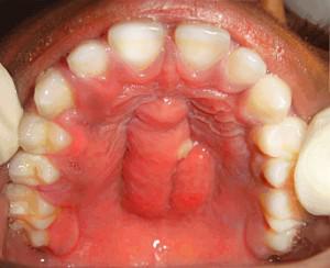
Periostitis of the lower jaw occurs against the background of a progressive purulent disease. In the lower jaw, pathological changes are possible not only in bone tissue, but also in soft tissue. Often untreated caries and advanced dental diseases are the cause of periostitis of the lower jaw. Odontogenic periostitis is a common form of inflammation, one of the symptoms of which is swelling, which can be seen in the photo below.
Periostitis occurs more often in the lower jaw than in the upper jaw. The first characteristic sign of this type of disease is a dull, increasing pain, which at the beginning of the disease manifests itself during eating or when pressing on a tooth, and swelling of the lower jaw area. Over time, the pain intensifies, radiates to the ear, temples, and swelling increases.
Dental periostitis often occurs among children who have dental problems due to advanced caries or pulpitis. The infection spreads through the blood and lymph. Ignoring the problem leads to aggravation of the situation and complications in the form of periostitis of the neck, eye sockets and other areas.

The inflammatory process in the periosteum can also occur due to decreased immunity and weakened protective function of the body, as a result of which odontogenic acute periostitis of the jaw develops. This disease is a recurrent disease, which after another exacerbation and relapse can develop into a chronic form. The disease may be a consequence of injury to this area, and the onset of aseptic (traumatic) inflammation of the jaw.
Other types of periostitis
The inflammatory process in the periosteum is possible not only on the jaw bones, but also on the heel, nasal, humerus, tibia, and fibula bones. Symptoms of the disease may also vary. There are the following types of disease:

Treatment of inflammation of the periosteum
Treatment of periostitis of the upper and lower jaw can be carried out depending on the situation, the size of the tumors, the severity and form of the disease using different methods. Often, dentists use several methods simultaneously to speed up the process of treating acute purulent periostitis and increase its effectiveness. The treatment method for jaw periostitis may be as follows:
- surgical (operative);
- therapeutic;
- medicinal;
- physiotherapeutic;
- unconventional.
During surgical intervention, the inflamed gum is opened and all contents and tissue affected by acute purulent periostitis are removed from the resulting cavity. Then the dental canals are opened, which are thoroughly cleaned of pus, after which the dentist treats them with medicine and installs a temporary filling. A few days later, at the next visit to the doctor, the canals are sealed and a permanent filling is installed on the tooth. To ensure the effectiveness of the treatment, the patient is given a control x-ray.
The therapeutic method involves opening the tooth, cleaning it of serous fluid and filling the canals. This method is effective only for acute serous inflammation of the periosteum.
In many cases, surgery is not necessary. The doctor prescribes a set of medications to the patient that will help stop the growth of gumboil, relieve inflammation and resist bacterial infection. Antibiotics, anti-inflammatory drugs, analgesics, and antihistamines are often prescribed. It is strictly not recommended to prescribe and take antibiotics on your own; this should be done by a doctor.
The physiotherapeutic method is most often used in cases of chronic and traumatic forms of dental periostitis. The essence of the method is to influence the tumor with devices such as a laser, UV lamp, electrophoresis and others.

Among the traditional methods of treatment, solutions and decoctions of herbs are used for rinsing. The most effective of them are a soda-salt solution, as well as a decoction of chamomile flowers, calendula, sage and other herbs that have antiseptic, wound-healing and anti-inflammatory properties. Warming up and compresses are strictly prohibited, as this will only aggravate the inflammatory process.
Complications of the disease
Untimely treatment of periostitis and not taking this problem seriously can cause a number of complications and complicate the treatment process. If you do not treat purulent periostitis, it can cost your life; at best, such a negligent attitude towards health will lead to the fact that the acute form will smoothly turn into chronic. The most harmless form of this disease is acute serous, in contrast to purulent periostitis, which poses a huge risk to human health and life.
In case of acute purulent periostitis, surgical intervention cannot be avoided, since not every flux can be opened without the help of a doctor. For example, if the abscess is located in the palate area, then opening it independently is impossible, and the lack of timely treatment is fraught with necrosis of the palatine bone and osteomyelitis.
The sooner qualified assistance is provided to the patient, the greater the chances of a successful outcome and a quick cure. You should not delay treatment, as the inflammatory process spreads rapidly and it becomes more difficult to cure.
Prevention of periostitis
Compliance with preventive measures will help prevent the occurrence of periostitis of the jaw and possible complications in the form of sepsis, osteomyelitis and other serious diseases.
Be sure to take proper care of your mouth and teeth. This will make it possible to avoid a number of problems - caries, stomatitis, pulpitis and purulent maxillary periostitis.
- To brush your teeth, you should use high-quality toothpaste, floss, a brush, and also use toothpicks and chewing gum if you cannot brush your teeth after every meal. After brushing your teeth, it is advisable to thoroughly rinse your mouth with a special product that removes what remains after brushing and fights pathogenic bacteria.
- Fear of dentists can cause dental diseases, including acute periostitis. It is necessary to visit the dentist at least twice a year, since regular examination by a doctor will help to identify the problem in time and eliminate it immediately, even if it is periostitis of a chronic form of the tibia.
- A balanced diet enriched with vitamins, beneficial and vital microelements is the key to the health of the whole body, including the dental part.
Periostitis, better known as “flux,” is an acute surgical pathology, purulent inflammation, which can be considered a threat to health, and if there is no timely treatment, it can even pose a threat to life. So what are the causes of flux, the symptoms, and why is it so dangerous?
Table of contents:What is periostitis?
Inflammation of the periosteum of the alveolar process of the jaw is called periostitis. If we consider statistical data, among all cases of acute inflammatory diseases of the maxillofacial area, “flux” is recorded in 6% of cases. In the vast majority of cases (96%), its course is acute; extremely rarely the process becomes chronic. You can also highlight the area where inflammation most often forms: 2 times more often due to diseases of the teeth of the lower jaw.

note
The popular term “flux” was not chosen by chance. In translation “Fluxus” means flow, flow. This value reflects what is happening - the spread of pus is comparable to a river during flood.
Periostitis is a complication of timely untreated caries complications. It cannot be said that periostitis is a dental disease, but it is impossible to deny that they are the cause. The pathology develops slowly: it passes into, then into, and then the inflammation spreads and the periosteum is involved in the process. This transition may take several years, but the lack of treatment, sooner or later, will come back with serious complications.
Typically, acute inflammation begins in the inner or outer layer of the periosteum, but gradually it spreads, which threatens the development of even greater problems.
Periostitis: types and classification
In clinical practice, a wide variety of types of periostitis can be distinguished, and their classification is quite extensive:
- by type of infection spread: odontogenic (infection from a tooth), with blood flow - hematogenous, lymphogenous (with lymph flow), traumatic (dental damage, tumor processes, wounds, etc.);
- along the course: acute, which in turn is divided into serous, purulent, diffuse, as well as chronic (simple and ossifying);
- by distribution: limited (inflammation involving 1-3 teeth), as well as diffuse (inflammation covers almost the entire jaw).
Each of these types has its own characteristics, which relate to the causes of their formation, symptoms, and treatment. In addition, the risks and number of possible complications will vary.
What are the causes of periostitis?
Statistics show that most often it is registered odontogenic periostitis, that is, resulting from complications of caries.
 If we consider the ways of spreading infection through blood and lymph, then the risks will be posed by diseases such as acute infections. But, as a rule, this route of spread is typical for children.
If speak about traumatic periostitis, then its cause may be hidden in a complex or atypical tooth extraction, serious injuries to the maxillofacial area, for example, jaw fractures, wounds, and others.
In addition to the main causes that directly affect the formation of acute inflammation, it is necessary to remember about predisposing causes and factors. We are not talking about brushing your teeth, as well as visiting the doctor on time, which is, of course, important. We are talking about hypothermia, untreated diseases of internal organs in a timely manner.
Symptoms of periostitis
Each form of periostitis has its own specific symptoms, but, nevertheless, there are also common ones:
- sharp pain spreading to the neck and head;
- significant swelling of the cheek, up to swelling of the eye;
- any chewing movement causes pain;
- in the oral cavity: redness of the gums, smoothing of the transitional fold;
- symptoms of intoxication.
It seems to patients that the symptoms of periostitis appear unexpectedly, but, in fact, “flux” has several stages of development:
- the appearance of pain in the tooth when chewing and pressing on it, which can also be considered a symptom of periodontitis;
- the gums around the causative tooth become brightly colored, after which it constantly grows and the transitional fold is smoothed out;
- Significant swelling of the cheek forms, sometimes spreading to the lip and wings of the nose. Everything will depend on the location of the causative tooth;
- a sharp increase in body temperature, the formation of a pulsating temperature, it can also spread to the ears, head and other areas.
All stages of development pass in just a few days.
In addition to general symptoms, each form has its own specific symptoms.
Acute serous periostitis
The appearance of acute symptoms in acute serous periostitis occurs 1-3 days from the onset of the process. The main symptom of this form can be considered the formation of facial edema. The location of the edema and, consequently, its size is directly dependent on the causative tooth.
note
This form of inflammation is more often associated with bruises and jaw injuries, and swelling and other acute symptoms disappear in just a few days.
Sometimes, in the presence of additional aggravating circumstances, acute serous periostitis can provoke fibrous growth, as well as deposition of calcifications in the periosteum. The acute serous form can become a predisposing factor for the transition of acute inflammation to chronic inflammation, namely periostitis ossificans.
Acute purulent periostitis
 This type of periostitis is characterized by the appearance of acute, unbearable throbbing pain. It is noteworthy that it can spread to the ear, temple, and eye. Typically, the pain appears in the evening or at night, causing patients to literally climb the wall. The relief may be short-lived, but then the pain comes back.
Afterwards, by the morning, serious swelling forms, and its increase occurs gradually: along with pain, swelling forms in the mouth, and the transitional fold is smoothed out. Then, it spreads to the cheek tissue.
note
Depending on the location of the tooth, swelling may spread to the eye area or to the neck area if the causative tooth is located on the lower jaw.
The main complaints of patients: deterioration in general well-being, decreased performance, increased temperature within 38.5º C, irritability.
The above symptoms may intensify as purulent discharge accumulates.
Acute diffuse periostitis
Symptoms of this form will be severe, widespread pain, spreading and radiating to nearby areas. General symptoms of intoxication also appear: poor health, headaches, fatigue, irritability, high body temperature (within 38.5º C), lack of appetite.
The course of acute diffuse periostitis of the lower and upper jaws is different. In the lower jaw, the disease is much more complicated, which can be explained by anatomical and physiological features.
As periostitis develops, edema forms, its localization and characteristics depend on the causative tooth:

Chronic periostitis
The main feature of chronic periostitis is the absence of clearly defined symptoms, in fact, the patient is not worried about anything. Fortunately, this form of periostitis is extremely rare and only in patients with serious diseases of the internal organs or problems with the functioning of the immune defense.
In the remission phase, few patients pay attention to the formation of dense swelling, which does not change facial features and also does not have any symptoms.
Chronic periostitis can exist and develop for a long time without making itself felt, literally for many months, or even years. But, sooner or later, an exacerbation will occur, and symptoms characteristic of the acute form will appear.
With exacerbation of chronic periostitis, the following symptoms often appear:
- spilled nature;
- swelling of the cheek;
- increased body temperature;
- poor health, loss of appetite.
Chronic ossifying periostitis
Periostitis ossificans is a fairly common form of chronic inflammation that leads to the formation of new bone. The formation of new bone usually occurs in a limited area that resembles spines; they can merge with each other and form dense tissue that varies in shape and size.
If we talk about symptoms, they boil down to:
- pain at night;
- dense swelling;
- may be accompanied by inflammation of the periosteum.
How is periostitis diagnosed?
Diagnosis of periostitis occurs in the dentist’s office, after analyzing the data obtained from a survey, examination and some types of research, including visual ones.
The first thing dentists start with is a survey. The doctor will be interested in when the symptoms appeared, what they were, whether the patient can point to a bad tooth, whether there were injuries and what is the general health. The main task of patients is to answer questions truthfully.
Afterwards, the doctor proceeds directly to an external examination: condition, size and density of edema, pain, condition and reaction of the lymph nodes. Only then does the dentist proceed to examine the oral cavity. Usually, identifying the causative tooth is not difficult: a large carious cavity, or the patient himself says that the tooth has previously been treated.
Dentists also identify characteristic signs:
- redness in the gum area and near the causative tooth;
- smoothing the transition fold;
- infiltrate;
- symptoms of fluctuation. The dentist presses on the swelling in the area of the causative tooth and a transfusion of inflammatory fluid is felt;
- crepetation. In some cases, when pressing on the gum in the area of the diseased tooth, a sound similar to the rustling of parchment is formed.
Dentists also conduct a number of tests:
- percussion - tapping on the tooth. This reaction causes sharp pain, the patient literally jumps in the dentist’s chair;
- probing - studying the depth of the carious cavity and its condition.
One of the main research methods that dentists use is. But at the same time, the pictures do not show any changes in the bone tissue, but x-ray signs of various forms of periodontitis, as well as dental cysts, can be detected.
Differential diagnosis
 In medicine, there is such a thing as differential diagnosis. Many diseases may have similar symptoms, but the causes, and even more so the treatment, differ significantly. Therefore, it is extremely important to distinguish one disease from another.
In the case of periostitis, the differential diagnosis is made between acute periodontitis, abscess formation, acute osteomyelitis, and also.
In addition to the general symptoms that are characteristic of all these diseases, you can find those that will prompt the correct diagnosis. The main thing is to notice them in time. In many ways, patients themselves help in the process of differential diagnosis.
How is periostitis of the jaw treated?
The treatment offered by dentists will depend on the stage of development of the process, the specific form of periostitis, the presence of complications and concomitant diseases.
Treatment in the early stages
In the early stages of the development of the process, the abscess is usually absent. Therefore, treatment can be carried out without surgery. A number of medications are selected for patients:
- painkillers;
- antipyretics;
- , as a rule, a wide spectrum of action;
- antiseptics for mouth rinsing.
Sometimes this treatment is not enough, and it is necessary to create an outflow of inflammatory fluid or pus. To do this, the dentist removes all tissues altered by caries, opens the root canals, thereby opening the passage for the release of accumulated pus and inflammatory fluid. During this time, patients are recommended to rinse their mouth with a soda-salt solution, and the more often they do this, the better.
The dentist also decides on an individual basis whether it is possible to save the tooth, or whether it would be more advisable to remove it.
note
If chronic inflammation has been diagnosed or an exacerbation has occurred again after conservative treatment, the root canals are impassable, and the patient has a history of a “bouquet” of chronic diseases, the tooth will most likely have to be removed.
Surgical methods of treatment
Surgical treatment can be carried out using the method of tooth preservation, as well as its removal. These are two different techniques, differing in the method of implementation, indications, as well as possible risks and complications.
Tooth-preserving surgery involves:
- opening the source of infection: the dentist dissects the mucous membrane, periosteum to allow the outflow of pus;
- assistance in the outflow of pus, washing the formed cavity, installing drainage, which is necessary to ensure that the outflow path of pus does not close;
- rinsing the mouth with soda-salt solutions to drain pus, as well as antiseptic treatment;
- rinsing the mouth with antiseptic solutions;
- taking broad-spectrum antibiotics in a course that must not be violated under any circumstances;
- taking anti-inflammatory drugs.
After the symptoms of acute inflammation disappear, the doctor continues treatment - restoration of bone tissue, stimulation of bone formation, opening and cleaning of root canals, and antiseptic treatment of the periodontal space are necessary. Then the doctor restores the damaged tooth crown.
If indications for tooth extraction have been determined, then the tooth is removed followed by treatment of the area of inflammation:
- curettage Removal of purulent contents of necrotic tissue;
- washing the hole of the extracted tooth with an antiseptic solution;
- taking antibiotics;
- laying down special medications that will help destroy the infection and prevent complications.
Patients after treatment of periostitis need close attention from dentists. After the operation, the dentist draws up a schedule of visits: the next day after the operation, on the 2nd, 3rd, 5th day of treatment. In the future, the visiting schedule is determined strictly individually.
How can you not treat periostitis?
Periostitis is a dangerous disease that can lead to serious illness and can be considered a threat to health and even life. Any delay, even the slightest, can be fatal.
Self-medication and “folk” remedies are something you should not do under any circumstances. The scope of “home” treatment includes:
- self-prescription of antibiotics. It is possible that acute inflammation can be stopped, but without further treatment, the “flux” will make itself felt again, and with even more pronounced symptoms;
- lack of treatment can cause inflammation to spread and develop into complications: abscesses of the head and neck. Let us recall that acute purulent processes and, especially, the accumulation of pus in the maxillofacial area, a place that is abundantly supplied with blood vessels, are dangerous for the spread of infection to the brain area;
- It is strictly forbidden to apply heat or use compresses. Such actions only accelerate the process of spreading pus, and the consequences will not be long in coming;
- reception . Taking this drug can also provoke the rapid development of complications.
Possible complications and prognosis of the disease
The accumulation of pus in the tooth area and inflammation of the periosteum is dangerous due to the development of numerous complications that can be considered as posing a threat to health and even life:
- cellulitis or soft tissue abscess: localized or diffuse purulent inflammation that can spread through the bloodstream to the brain area or lower to the heart area. The spread of purulent inflammation is dangerous due to the spread of infection throughout the body and the development of sepsis;
- osteomyelitis of the jaw– purulent inflammation of the bone, which will threaten the development of inflammation, as well as literally melting the jaw bone. This pathology is extremely difficult to treat and requires significant surgical intervention, transplantation operations, and the use of serious medications.
note
If we talk about prognosis, then acute serous periostitis of the jaw proceeds most favorably. It is difficult to say that the course of acute purulent periostitis and its prognosis will be difficult; the main thing is to intervene in a timely manner.
Prevention of periostitis
Knowing the main reason for the development of periostitis - dental disease, all preventive measures will be aimed specifically at their timely treatment and sanitation of the oral cavity.
- satisfactory oral hygiene. Hygiene products and items must be selected individually. Also, at the appointment, the doctor can give specific recommendations for brushing your teeth;
- professional oral hygiene in the dentist's chair: polishing the enamel, saturating the enamel with minerals;
- regular preventive examinations in the dentist's chair for timely treatment of caries in order to prevent its complications;
- sanitation of the oral cavity: healing of carious teeth, caries complications, inflammatory and inflammatory-dystrophic diseases of the oral cavity;
- injury prevention.
To prevent the formation of an acute inflammatory process in the periosteum, it is enough to regularly visit the dentist for the timely treatment of caries. It is important to prevent its complications. Only in this case can all risks be prevented.
Periostitis is an inflammation of the periosteum, which is.
In the absence of treatment for inflammation of the periodontium, the ligamentous apparatus of the tooth, the infection penetrates through the periodontal gap and affects the periosteum.
Types of disease
Depending on the intensity of inflammation, the following courses of periostitis of the jaw are distinguished:
- spicy;
- chronic.
Classification occurs based on the participation of pathogenic microflora in the inflammatory process:
- purulent;
- aseptic.
According to the nature of the exudate, periostitis occurs:
- exudative;
- proliferative (virtually without the formation of exudate).
The form of the disease is:
- simple;
- serous;
- fibrous;
- ossifying;
- otogenic;
- hematogenous;
- purulent.

Acute and chronic form of the disease
The acute form of periostitis is purulent inflammation of the periosteum of the jaw. Most often, the periosteum of the alveolar process becomes inflamed. In rare cases, an abscess of the palatine or sublingual fold is diagnosed.
The cause of the development of pathological processes in the lower jaw is often the molars. In case of acute inflammation of the upper jaw, the infection penetrates through the incisors, fangs, .
Chronic periostitis is rare. Inflammatory processes most often occur in the lower jaw. This pathology is observed in patients with immunodeficiency conditions.
After acute inflammation and pain subsides, the abscess may spontaneously empty. As a result of chronic recurrent processes, bone compaction remains. The chronic course is observed more often in adolescents and children.
Causes and risk factors
Purulent acute processes arise as a result of the penetration of streptococcal and staphylococcal infections. Acute inflammation is a complication of recurrent periodontal inflammation.
Pathology can develop against the background of:

Unfavorable contributing factors include:
- hypothermia;
- stress;
- fatigue;
- injuries;
- decreased immunity.
In acute and recurrent chronic periodontitis, pus is not able to freely penetrate outward. In this regard, the contents of the abscess begin to penetrate through the lymphatic vessels into the periosteum.
With this violation, the integrity of the tissue is compromised. This determines the insufficiency of cellular immunity to fight infection.
Features of the clinical picture
As inflammation develops, the periosteum thickens due to swelling and detachment from the bone. Hemorrhages may form in the affected area due to vascular spasm. There are small infiltrates around the periosteum, and swelling of the connective tissue is observed.
During the purulent process, the inner surface of the periosteum begins to quickly melt. Exudate begins to accumulate between the bone and the inner layer.
Symptoms of acute periostitis
As a result of local immune reactions, small abscesses begin to appear, which eventually merge with each other.
A large mass of pus contributes to even greater detachment of the periosteum.
The result of periosteal necrosis is a violation of the integrity of the exfoliated periosteum and a breakthrough of the contents under the mucous membrane of the oral cavity.
In the area of suppuration, small vessels are destroyed and thrombosed. After a few days, the abscess may break into the oral cavity.
The symptoms of a purulent process depend on the location and duration of the inflammatory process.
The symptoms of acute periostitis of the jaw are multifaceted:
- Patients complain about pain, swelling in the area of inflammation.
- During acute inflammation temperature rises, weakness and malaise appear. At the beginning of inflammatory processes, the swelling is small. Within two days, symptoms rapidly intensify.
- In the patient Pain appears throughout the jaw, eye area, ear, temple. After thermal procedures, inflammation worsens. The cold helps the pain subside for a while.
- Patients note that First there is pain in the tooth, and later facial swelling occurs in the affected area. After swelling appears, the intensity of the pain symptom decreases.
Acute purulent periostitis of the jaw has its own symptoms:
- With purulent inflammation of the jaw in the affected area swelling appears. When the upper incisor becomes inflamed, the upper lip and wings of the nose swell.
- When the infection spreads swelling covers a large area of the face. When the molars in the upper jaw become inflamed, the zygomatic and parotid areas, cheek, and lower eyelid swell.
In acute periostitis, which occurs as a result of inflammation of the lower incisors, the lower lip and chin swell. The spread of infection contributes to swelling of the lower cheeks and corners of the mouth.
When the molars in the lower jaw become inflamed, the cheek, parotid and submandibular areas swell. In this case, patients experience severe pain and limited joint mobility due to muscle contracture.
The acute process is accompanied by regional inflammation of the lymphatic vessels, swelling of the mucous membrane in the area of the affected tooth, and a painful infiltrate.
After the abscess breaks through into the submucosa, the pain subsides and swelling decreases. Pus is often visible through the mucous membrane of the mouth. Sometimes the abscess itself breaks into the oral cavity. After the pus is released, the inflammation subsides.
When the lingual surface of the alveolar process in the lower jaw is inflamed, patients complain of pain when swallowing, moving the tongue and jaw.
On examination, redness and swelling of the palate are noted. If the alveolar process of the upper jaw becomes inflamed, a palatal abscess occurs. In patients, the submandibular lymph nodes are enlarged.
As the abscess enlarges, the folds in the palate are smoothed out. When the infection spreads, the soft palate and velopharyngeal arch are affected.
Patients experience throbbing pain that gets worse when moving the jaw. After a week, the pus may break into the oral cavity on its own.
Signs of a chronic disorder
With chronic inflammation, the periosteum increases due to the growth of connective tissue. Depending on the age of the patients and  state of immunity, the formation of cat tissue is observed at different stages of maturation. First, coarse fibrous plates are formed, later lamellar bone is formed.
The disease has a long course with periods of exacerbation. The patient may suffer from chronic periostitis for several years.
During the examination, the dentist discovers minor changes in the structure of the face, notes a painful compaction of the bones and lymph nodes in the submandibular region. In the oral cavity, swollen, reddened mucous membranes are noted.
Treatment of the disease
In case of acute periostitis, complex treatment is carried out. The patient is opened an abscess, after which conservative therapy is prescribed.
In the initial stage of inflammation (serous inflammation), treatment begins with creating conditions for the outflow of exudate. The patient's tooth cavity is opened and decay products are removed from the canals.
In more advanced cases, the patient who was the cause of the development of periostitis. The doctor performs all manipulations under local anesthesia (). Usually after such events the inflammatory process stops.
In case of acute periostitis of the upper jaw, urgent surgical care is indicated. Patients are opened a purulent focus, the wound is treated and  create conditions for the outflow of abscess contents. Such operations are usually performed in the department of maxillofacial surgery under local anesthesia. After opening the abscess, a rubber drainage is inserted into the wound to drain the exudate.
In the postoperative period, rinsing the mouth with antiseptics and washing the wound with Ethacridine, a solution of Dimexide with Oxacillin are indicated.
Treatment with drugs consists of prescribing:
- nitrofuran agents (Furadonin);
- sulfonamide drugs (Sulfadimezin);
- antibiotics (Oletetrin, Oxacillin, Lincomycin);
- painkillers;
- antihistamines (Suprastin);
- vitamins
After removal of the abscess and subsidence of acute inflammation, the patient is shown physiotherapeutic procedures:
- Sollux lamp;
- laser therapy;
For chronic inflammation, treatment consists of removing the source of infection and prescribing physiotherapeutic procedures. If the disease lasts for a long time and treatment is ineffective, bone formations are removed.
Danger is near
As a result of pathological processes, the cortical layer of the jaw becomes thinner, which can lead to the formation of defects. Sometimes, with severe detachment of the periosteum, a secondary one occurs with massive infiltration.
If the abscess under the periosteum is located on the periphery, a periosteal neoplasm may develop. If there are errors in treatment and failure to follow the doctor’s recommendations, the infection can spread to the bone and lead to osteomyelitis of the jaw, phlegmon or abscess.
Preventive actions
Prevention of acute periostitis involves timely treatment of odontogenic inflammation. To prevent chronic forms  pathology should be combined with preventive measures, correction of immunity.
Timely treatment of acute periostitis results in complete recovery. Already on the fifth day, patients become able to work.
In chronic processes, as a result of adequate therapy, recovery occurs. In advanced cases, patients may develop chronic cortical osteomyelitis.
5.2. PERIOSTITIS
Periostitis - This is a disease that is characterized by the spread of the inflammatory process to the periosteum of the alveolar process and the body of the jaw from an odontogenic or non-odontogenic focus.
Periostitis of the jaws occurs in 5.2-5.4% of patients undergoing treatment in the clinic (Ya.M. Biberman, 1965; A.N. Fokina, D.S. Sagatbaev, 1967). Among patients with odontogenic inflammatory processes of the jaws, periostitis was treated on an outpatient basis in 3.42% and in a hospital in 19.17% (Mauks, 1975). According to our data (A.A. Timofeev, 1983), 20-23% of patients who were hospitalized for inflammatory diseases experienced periostitis, mainly its acute form (in 94% of patients).
Periostitis was localized on one side of the jaw, most often affecting it from the vestibular surface (in 93.4% of patients). In the area of the lower jaw, periostitis was observed in 58.9% of patients, in the upper jaw - in 41.1% (G. Vasiliev, T. G. Robustova, 1981), and according to our data - in 61.3 and 38.7%, respectively. (A.A. Timofeev, 1983).
♦ Acute periostitis
The occurrence of acute odontogenic periostitis is preceded by the following diseases: exacerbation of chronic periodontitis - in 73.3% of patients; alveolitis - in 18.3%; difficult eruption of wisdom teeth - in 5.0%; suppurating odontogenic cysts of the jaws - in 1.7%, periodontitis - in 1.7% of patients. The disease most often develops after a traumatic tooth extraction operation, with incomplete tooth extraction, and less often after an atraumatically performed surgical intervention. Trauma associated with tooth extraction can cause activation of a dormant infection located in the periodontal gap, which leads to the spread of the inflammatory process under the periosteum.
Acute odontogenic periostitis comes in serous and purulent forms. Serous periostitis is considered as a reactive inflammatory process in the periosteum, which accompanies aggravated chronic periodontitis. With purulent periostitis, exudate from the inflamed periodontium penetrates under the periosteum through the Volkmann and Haversian canals, through the lymphatic vessels or through a previously formed hole in the wall of the socket (GA. Vasiliev, 1972).
I believe that with such a mechanism of spread of the infectious process, it is difficult to imagine acute odontogenic periostitis, complicated by abscesses and phlegmons, occurring without pronounced destruction of bone tissue. In an experiment conducted on animals and human corpses, which consisted of introducing a methylene blue solution into the tooth socket, root canal or periodontal fissure under pressure, A.I. Vasilenko (1966) noted its spread along the medullary beams to all parts of the lower jaw and surrounding soft tissues.
According to M.M. Solovyov and I. Khudoyarov (1979), during periostitis, the spread of the infectious process under the periosteum by the lymphogenous route is less likely, since in these cases one can more likely expect retention of microbes, toxins and tissue decay products in the regional lymph nodes and the subsequent development of lymphadenitis and adenophlegmon. The authors believe that the formation of an abscess in the perimaxillary soft tissues is not associated with the breakthrough of pus under the periosteum, but with the formation of “own” pus in this place under the influence of microorganisms, bacterial toxins and tissue decay products.
In my opinion, the products of tissue decay of microorganisms, toxins, and sometimes the microbes themselves from odontogenic foci penetrate into the periosteum along the vessels that pass in the canals of the compact layer of bone. The first penetration of these substances usually does not cause the development of an inflammatory process, but only forms local sensitization of tissues. The subsequent entry of microbes into the body, as well as a decrease in its reactivity, with an increase in allergenicity, in paraallergic reactions (hypothermia, overheating, physical stress, etc.) causes the development of infectious-allergic inflammation with subsequent effusion of exudate under the periosteum of the jaw (A.A. Timofeev, 1982).
It has been established that the causative agent of the disease is usually non-pathogenic staphylococcus. Since the waste products of this microflora do not have a damaging effect, the author assigns a special role to allergy mechanisms in the occurrence of odontogenic periostitis.
When studying, using intradermal tests and laboratory tests, the microbial sensitization of the patient’s body to pathogens located in the focus of purulent inflammation of the jaw, we found that in patients with acute odontogenic periostitis it occurs in response to the action of certain bacterial allergens. The body's sensitization to the staphylococcus allergen was 3 times higher than normal, and to the streptococcus allergen - 2 times (A.A. Timofeev, 1982). In the occurrence of acute odontogenic periostitis of the jaws, the main predisposing factor is microbial sensitization to staphylococcus, the frequency and severity of which correlates with the severity and prevalence of the process. In case of uncomplicated acute odontogenic periostitis, we registered it in 22% of patients, and in case of complications of its course by purulent processes in the peri-maxillary soft tissues - in 46%.
Thus, the participation of allergy mechanisms explains the reasons for the development of odontogenic inflammation caused by non-pathogenic microflora, and the predominant damage to the perimaxillary soft tissues that occurs in certain forms of acute odontogenic inflammatory diseases (M.M. Solovyov, I. Khudoyarov, 1979).
CLINICAL PICTURE . Clinical manifestations during acute odontogenic periostitis of the jaws are varied and largely depend on the general and local reactivity of the patient’s body, the type of inflammatory reaction, the virulence of the microflora and the localization of the inflammatory process in the periodontium and the age of the patient. In most cases, it is possible to establish a connection between the occurrence of periostitis and previous paraallergic reactions: hypothermia, overheating, physical or emotional stress. In other patients, especially those with reduced body reactivity, the disease develops more slowly. This course of the process is especially often observed in elderly and senile people, as well as in the presence of concomitant diseases, such as diabetes mellitus, circulatory disorders of II-III degree, chronic diseases of the digestive system.
According to our research, the cause of acute periostitis of the lower jaw in 22.9% of patients is a focus of inflammation located in the tissues of the first large molars, in 17.8% - the third large molars, in 12.3% - the second small molars. The development of acute odontogenic periostitis of the upper jaw is caused in 24.8% of patients by the presence of a focus of inflammation in the tissues of the first large molars, in 11.6% - in the second large molars (A.A. Timofeev, 1982).
In acute periostitis, the inflammatory process develops vestibularly in 93.4% of patients and occurs in an acute serous form in 41.7%, in an acute purulent form in 58.3%. With this form of acute odontogenic periostitis, detachment of the periosteum over 1 tooth is noted in 20% of patients, over 2 teeth - in 56%, over 3-4 teeth - in 24%.
Patients complain of pain in the tooth, which intensifies when touching it with the tongue or antagonist tooth, and swelling of the face. The pain, which was previously localized in the area of the causative tooth, during this period is characterized as pain in the jaw. In 8.9% of patients, irradiation was noted along the branches of the trigeminal nerve to the area of the ear, temple, and eye. The general condition of patients worsens, weakness, headache, sleep disturbance, loss of appetite, chills, and malaise appear.
Paresthesia of the lower lip (Vincent's symptom) is noted in 11.7% of patients and is determined only in those in whom the inflammatory process is localized in the lower jaw in the area of large and small molars.
Body temperature increases in 92% of patients: in 20% - from 37 to 37.5 ° C, in 28% - from 37.6 to 38 ° C, in 44% - from 38, HS and above.
In acute odontogenic periostitis of the jaws, soft tissue swelling appears, which can be expressed to varying degrees. The localization of edema is usually quite typical and depends on the location of the causative tooth. At the onset of the disease, soft tissue swelling is most pronounced. According to V.G. Lukyanova (1972), the amount of edema depends on the structure of the vascular (venous) network of the periosteum. With the finely looped form of branching of the vessels of the lower jaw, the swelling of the soft tissues is little pronounced, while with the main form (the area of the tubercle of the upper jaw, the angle and branches of the lower jaw) it has a significant extent. We observed pronounced swelling of the soft tissues around the affected area in 67% of patients with acute odontogenic periostitis of the upper and lower jaws.
Upon palpation of the soft tissues at the location of the subperiosteal inflammatory focus, a dense, painful infiltrate was determined. In 82% of patients, regional lymph nodes were painful, enlarged, had a dense elastic consistency, but remained mobile. When the inflammatory process was localized in the area of large molars, 60% of patients with acute periostitis experienced inflammatory contracture muscles:I- when there is only a slight restriction in mouth opening;II- when the mouth opens 1 cm;III- when the jaw is tightly closed and opening the mouth independently is impossible. In other cases, the restriction of mouth opening is associated with the fear of opening the mouth wide due to the pain that arises.
When examining the oral cavity in the area of the affected teeth, one can detect hyperemia and swelling of the mucous membrane of the transitional fold and the alveolar process of the jaw. As a result of examination of patients with acute odontogenic periostitis of the jaws, its serous form was revealed in 42%, and purulent in 58%. When the process passes into a purulent form, a roller-like protrusion is formed along the transitional fold - a subperiosteal abscess. If pus melts the periosteum and spreads under the mucous membrane, it forms subdess neve (submucosal) abscess.
The crown part of the causative tooth is partially or completely destroyed, the carious cavity and root canals are filled with putrefactive contents. A deep periodontal pocket may be found in the area of the tooth that served as the source of infection. Sometimes this tooth is filled. A painful reaction to percussion of the causative tooth was noted in 85% of those examined, and of neighboring teeth (one or two) - in 30%. The causative tooth becomes mobile in 37% of patients. When acute periostitis occurred as a result of alveolitis in 10% of patients, we observed (within 2-3 days) the release of purulent exudate from the socket of the extracted tooth. In 60% of patients with odontogenic periostitis, acute sinusitis was detected, which was a complication of the inflammatory process in the upper jaw when localized in the area of large and small molars (A.A. Timofeev, 1982).
An X-ray examination of the jaws did not reveal changes characteristic of acute periostitis; granulating or granulomatous periodontitis, perihilar cysts, semi-impacted teeth, etc., which preceded the process, were found (Fig. 5.1.1-5.1.5).
No changes in the phagocytic activity of peripheral blood neutrophil granulocytes were detected in patients (the exception was patients with concomitant diseases). The results of a blood test during the initial period of disease development indicate an increase in the number of leukocytes (9-12 * 10 9 / l), and sometimes higher. Only in some patients the number of leukocytes is within normal limits or leukopenia is observed. The increase in the number of leukocytes occurs due to segmented neutrophilic granulocytes (70-76%) and their band forms (8-20%). The number of eosinophilic leukocytes can decrease to 1%, and lymphocytes - to 10-15%. ESR increased to 19-28 mm/h, and sometimes more. In patients with acute odontogenic periostitis, a 2-4 times increase (compared to healthy people) in the activity of alkaline and acid phosphatases of neutrophil granulocytes in peripheral blood was detected. In most patients, no changes were found in urine tests; only in some people with high body temperature, protein appeared in the urine (from traces to 0.33 g/l), and sometimes leukocytes.
When studying microbial sensitization of the body, its presence was established in 22% of patients with acute odontogenic periostitis and in 46% when it was complicated by purulent processes in the perimaxillary soft tissues. The body's sensitization to the staphylococcus allergen increased 3 times, and to the streptococcus allergen - 2 times. The presence of the fact of preliminary microbial sensitization subsequently served as the basis for nonspecific microbial hyposensitization in patients with this disease (A.A. Timofeev, 1982).
Features of the clinical course of acute odontogenic periostitis depending on the localization of the process . The clinical course of acute odontogenic periostitis depends on the location of the tooth that caused the development of the inflammatory process.
When the inflammatory process spreads from the source, located on the upper jaw on the vestibular side, in the area of the incisors there is significant swelling of the upper lip and wing of the nose, which can spread to the bottom of the lower nasal passage. In some cases, purulent exudate can penetrate under the periosteum of the anterior part of the bottom of the nasal cavity, especially with a low alveolar process, and form an abscess there. In the case when the inflammatory process begins from a focus located in the area of the central incisor, the swelling can spread to the entire upper lip, and if in the area of the lateral incisor, it can involve the soft tissues of one half of the face. When purulent exudate spreads from the lateral incisor towards the hard palate, in the area of its anterior section, a hemispherical or oval-shaped swelling, painful when touched, appears and a palatal abscess is formed.
In cases where the cause of the disease is an inflammatory process, located in the area of the upper canines, swelling spreads to the infraorbital and part of the buccal region, the corner of the mouth, the wing of the nose, the lower and even the upper eyelid. The source of inflammation is always located on the vestibular surface of the alveolar process of the upper jaw.
If the source of infection is an inflammatory focus, located in the tissues of the small molars of the upper jaw, then the collateral edema covers a significant area of the face and is located somewhat to the side. It spreads to the infraorbital, buccal and zygomatic areas, often to the lower and upper eyelids. The nasolabial fold is smoothed out, and the corner of the mouth is lowered. Facial swelling may be absent when purulent exudate from the palatal roots of 414 teeth has spread to the palatal surface. In this case, a hemispherical protrusion is formed in the middle part of the hard palate - a palatal abscess. Constant contact of the palatal abscess with the tongue causes increased pain, so the patient’s eating and speech are difficult.
Acute odontogenic periostitis, arising from a source of inflammation located in the area of the upper molars, is characterized by swelling involving the zygomatic, buccal and upper part of the parotid-masticatory areas. Swelling rarely spreads to the lower eyelid, and almost never spreads to the upper eyelid. The swelling reaches the ear. A few days after the development of the process, swelling of the soft tissues begins to descend downwards, which can create a false impression that the pathological focus comes from the small and large molars of the lower jaw. When purulent exudate spreads from the palatal root of the 616 teeth towards the palate, there is no facial asymmetry. Detachment of the dense periosteum in this area causes severe aching and then throbbing pain in the palate. Due to the fact that there is no submucosal layer on the hard palate, the swelling is insignificant. Spontaneous opening of the abscess usually occurs on the 7-10th day, which can lead to the development of cortical osteomyelitis.
For purulent periostitis, in which the inflammatory process spreads from tissues to the area of the lower incisors, Characteristic is the presence of swelling of the lower lip, chin and mental area. At the same time, the chin-labial groove is smoothed out. When the inflammatory process spreads to the periosteum from a focus located in the area lower canine and small molars, swelling affects the lower or middle parts of the buccal region, the corner of the mouth and spreads to the submandibular region. If the source of infection is a focus of inflammation located in large molars of the lower jaw, then collateral edema affects the lower and middle parts of the buccal region, the parotid-masticatory and submandibular regions. When the inflammatory process spreads to the periosteum in the area of the angle and branch of the lower jaw, the swelling is not clearly expressed, but has a significant extent. Due to the fact that the masticatory muscles are located here, inflammatory contracture appears.
Examination of the lymph nodes in acute purulent periostitis, especially when the process is localized in the lower jaw, allows us to note that not just individual nodes, but entire groups of them are enlarged and painful.
Anatomically, in the lower jaw, the inner bone wall is thinner than the outer one. Therefore, acute periostitis, the cause of which was a lesion located in the area of large molar lower teeth, may spread to the lingual surface of the alveolar process. In this case, hyperemia and swelling of the mucous membrane of the alveolar process and sublingual region are observed. The hyoid ridge on the affected side enlarges and bulges between the tongue and the lower jaw. The tongue is swollen, coated, teeth marks are visible on it, its movements are painful, it is raised and shifted to the healthy side. If the inflammatory process spreads from the lower wisdom teeth, then the infiltrate may be located in the area of the pterygomandibular fold and the anterior palatine arch, which causes severe pain when swallowing. When inflammation invades the pterygoid muscles, inflammatory contracture occurs.
These are the main signs of the clinical manifestation of acute odontogenic periostitis. It should be emphasized that most of them are also inherent in other acute inflammatory diseases of the jaws, so differential diagnosis is necessary.
Pathomorphological changes in periostitis of the jaw characterized by the accumulation of purulent exudate between the bone and periosteum. Dystrophic changes occur in bone tissue: lacunar resorption of bone substance, expansion of the Haversian canals and medullary spaces. As a result of these processes, significant thinning occurs and, in some areas, disappearance of the cortical bone layer and adjacent bone beams. At the same time, penetration of purulent exudate from under the periosteum into the Haversian canals and its transition to the peripheral areas of the bone marrow spaces is noted (GA. Vasiliev, 1973).
DIAGNOSTICS . Differences between acute (exacerbated chronic) periodontitis from acute odontogenic periostitis are determined by the fact that in the first case, the source of inflammation is localized within one tooth, and in the second, the inflammatory process goes beyond its boundaries and spreads to the periosteum. The course of acute odontogenic periostitis is characterized by such signs as chills, facial asymmetry, thickening of the alveolar process, mobility of the causative tooth, positive percussion and mobility of adjacent teeth, inflammatory contracture of the jaws (when the process is localized in the area of large molars of the lower jaw). Using laboratory methods, it has been proven that in patients with acute odontogenic periostitis, the activity of neutrophil granulocyte phosphatases in the early phase of inflammation is significantly increased (A.A. Timofeev, 1981).
In the clinical picture of acute odontogenic periostitis, uncomplicated and complicated by perimandibular purulent processes, we did not identify a significant difference in the frequency of occurrence of clinical symptoms, which can be used to make a differential diagnosis of this disease and acute odontogenic osteomyelitis of the jaws in the early phase of inflammation. It follows from the above that carrying out their early diagnosis only based on individual clinical symptoms has great difficulties and is based on the totality of clinical data. (A.A. Timofeev, 1982).
Acute odontogenic periostitis should be differentiated from inflammatory diseases of the sublingual and submandibular glands and their ducts. It should be remembered that with periostitis, the salivary glands are never involved in the inflammatory process. When massaging the inflamed salivary glands and their ducts, muddy or purulent-streaked saliva is released from the mouths of the excretory ducts. With the help of radiographic examination of the soft tissues of the floor of the mouth, salivary stones can be detected in the bite (with calculous sialadenitis).
When carrying out differential diagnosis of acute odontogenic periostitis and acute nonodontogenic lymphadenitis it is necessary to examine the alveolar process of the jaw. With lymphadenitis of non-odontogenic origin, there are no changes in the teeth and mucous membrane of the alveolar process of the jaw, which occur in acute periostitis.
TREATMENT . In acute serous odontogenic periostitis, removal of the causative tooth leads to recovery. The subsidence of inflammatory phenomena is facilitated by the appointment of physiotherapeutic methods of treatment (UHF in an athermic dose, warming compresses, Dubrovin bandages, fluctuarization, helium-neon laser rays, etc.).
In case of acute purulent periostitis, the tooth that served as the source of infection is removed if it is of no functional or cosmetic value. In other cases, it is preserved and after the inflammatory process subsides, it is subject to treatment. Simultaneously with tooth extraction, the subperiosteal abscess is opened. An incision is made along three teeth, cutting through the mucous membrane and periosteum along the transitional fold to the bone. To prevent the edges of the wound from sticking together, it is drained. The drainage is left in the wound for 1-2 days. After opening the abscess on the hard palate, a small section of soft tissue (triangular in shape) is excised. This prevents the edges of the wound from sticking together and ensures reliable drainage of the palate abscess. In the case where the inflammatory focus is located on the lingual side of the lower jaw, the abscess is opened with a linear incision, which is made above the transition point of the mucous membrane of the alveolar process into the sublingual region.
In the postoperative period, patients are prescribed drug treatment: antibiotic therapy - prescribed only to weakened persons or with concomitant diseases (ampicillin or oxacillin sodium salt, oleandomycin phosphate or oletethrin, monomycin, kanamycin), sulfonamide drugs - sulfadimethoxine, sulfapyridazine; painkillers (amidopyrine, analgin, phenacytin or paracetamol); sleeping pills.
Due to the fact that in patients with acute odontogenic periostitis of the jaws, the presence of preliminary microbial sensitization was revealed, which can be eliminated by conducting a course of nonspecific hyposensitizing therapy, we prescribed the following drugs to all of them: diphenhydramine, diazolin, suprastin, etc.
To remove pus secreted from the wound into the oral cavity and to quickly resolve the inflammatory infiltrate, mouth rinses (40-42X) with a warm weak solution of potassium permanganate, 1-2% sodium bicarbonate solution or furatsilin solution (1:5000) 3-4 were prescribed once a day. For antiseptic rinses, you can use an infusion of chamomile flowers, calendula, sage leaves and other medicinal plants. I.G. Lukomsky (1955) believes that the movement of a warm mass of solution in the oral cavity (when rinsing) is an effective hydrotherapeutic procedure that promotes the rapid elimination of inflammation. The use of higher temperature solutions for these purposes causes stagnation in the inflammation site (Yu.I. Vernadsky et al., 1983).
Particular attention was paid to the treatment of regional lymphadenitis, since this complication can develop into an independently existing disease and thereby significantly prolong the period of disability of patients. On the 2nd - 3rd day, patients were prescribed UHF therapy at an athermic dose and fluctuarization. To treat lymphadenitis, you can also use warming semi-alcohol compresses at night, electrophoresis with potassium iodide, magnetic applicators, and helium-neon laser beams.
In order to prevent acute sinusitis, which can occur as a complication of odontogenic periostitis of premolars and molars of the upper jaw, it is necessary to prescribe daily use (for 5-6 days) of vasoconstrictor drugs (1-3% ephedrine solution, 0.1% solutions of naphthyzine or sanorin , galazolin) and UHF or microwave to the area of the maxillary sinus. To make sure that acute sinusitis has not taken a chronic course, after 2 weeks you can conduct a control x-ray examination of the paranasal cavities.
♦ Chronic periostitis
In adults, the disease develops rarely and, according to our data, occurs in 5.3-6% of patients with periostitis (V.V. Roginsky et al., 1983). The pathological process occurs more often in young people or children, and is often localized in the lower jaw. (V.G. Lukyanov, 1972). Distinguish simple and ossifying chronic periostitis, as well as his reifying form. At simple chronic periostitis the newly formed osteoid tissue undergoes reverse development after treatment, with ossifying form - Bone ossification develops in the early stages of the disease and most often ends in hyperostosis. Referential periostitis characterized by pronounced resorptive phenomena and restructuring of bone structures.
The cause of chronic periostitis of the jaws, As a rule, it is a transition from the acute form of the disease. Preceded by chronic periodontitis and trauma. The disease can occur due to suppuration of jaw cysts, inflammatory processes in the maxillary sinuses, as well as as a result of trauma caused by removable and fixed dentures. The presence of a chronic inflammatory focus in the periodontium causes in some patients a sluggish current localized inflammation of the periosteum with a predominance of the productive component (Ya.M. Biberman, A.G. Shargorodsky, 1985). Since chronic periostitis is not always preceded by an acute phase of the process, it should be classified as a primary chronic disease. At pathological examination it is clear that the affected area of the periosteum is a spongy bone tissue, on the surface of which there is a thin cortical layer. The network of interwoven bone trabeculae has varying degrees of maturity - from osteoid beams and primitive coarse-fibrous trabeculae to mature lamellar bone tissue. The bone tissue found in these layers is also at different stages of maturation (Ya.I. Gutner, N.I. Kushnir, 1970). Chronic proliferative inflammatory changes in the periosteum are difficult to reverse or cannot be reversed at all.
Severe toothache, swelling of the gums, gumboil and distortion of the facial contour are obvious symptoms of periostitis - an inflammatory disease of the periosteum or jaw bone, which affects approximately 20% of patients in dental clinics.
Without timely treatment, periostitis progresses, pain intensifies, and the purulent tumor increases. Patients have a high temperature, the infection spreads to neighboring tissues. In the most severe cases, blood poisoning and death can occur. How to recognize periostitis in time and prevent complications - read this article.
Symptoms of periostitis
Manifestations of periostitis differ depending on the patient’s age, stage and form of the disease, and individual characteristics of the body. However, in most cases there are a number of obvious signs indicating the disease:
- toothache - can “radiate” to the ear, eye socket and/or temple, sometimes the pain does not allow you to open your mouth normally and limits jaw movements;
- pathological tooth mobility;
- swelling of the gums, lips - in some cases, swelling distorts the face;
- hyperemia of the oral mucosa (overflow of blood vessels);
- the appearance of an abscess - a purulent tumor, which is popularly called gumboil;
- enlargement and thickening of lymph nodes in the face and neck;
- feeling unwell - body temperature within 38 degrees, weakness, headache, etc.
Sometimes a fistula tract forms in the flux, through which pus enters the oral cavity. Painful symptoms when such a course appears become less pronounced.
Unlike pulpitis and periodontitis, which are also accompanied by pain, with periostitis there is always a distortion of the contours of the face. Mildly expressed or very noticeable deformations are always present.
Doctors note that in most patients the disease makes itself felt after physical or emotional stress, hypothermia or overheating.

When making a diagnosis, doctors pay attention to symptoms. Methods such as:
- visual inspection;
- radiography;
- laboratory blood test.
A visual examination may reveal a tooth affected by caries with the crown part destroyed by more than 50%. It has a pronounced carious cavity, the canals are infected and filled with decay products of the pulp - neurovascular tissue. In 85% of all cases, such teeth react painfully to percussion - tapping.
An X-ray of a patient's tooth with periostitis most often shows periodontitis (inflammation of the tissue between the root and the jaw bone), cysts, impacted (unerupted) teeth, and newly formed bone tissue.
A blood test shows a slight increase in white blood cells. The ESR also increases slightly - no more than 12-15 mm/h.
When diagnosing, it is important to distinguish periostitis from osteomyelitis of the jaw and manifestations of specific inflammatory processes (syphilis, tuberculosis). In the first case, the images will show pronounced bone destruction, which is not typical for periostitis. In the second, there will be no sharp acute pain or swelling.

Classification of periostitis
According to ICD 10, periostitis is an inflammatory disease of the jaw. Like any inflammatory disease, it has different forms and stages.
Due to the occurrence
- Odontogenic - a consequence of untreated dental diseases (pulpitis, periodontitis);
- hematogenous - caused by infection entering the blood and its further spread;
- lymphogenous - a complication from the entry of pathogenic flora into the lymphatic system;
- traumatic - the result of mechanical damage to the periosteum, jaw fracture, surgical manipulation.
By degree of distribution
Depending on the degree of spread of infection, limited and diffuse periostitis are distinguished. The first affects the area of one or several teeth, the second affects a large group of teeth or the entire jaw.
By localization
In 61% of cases, periostitis of the lower jaw is observed, and only in 39% the infection is localized in the periosteum or bone of the upper jaw.
According to the course of the disease
- Acute - it accounts for 95% of all cases;
- chronic - 5% of cases.
Acute periostitis can occur in two forms - serous (41% of all patients) and purulent (59%). With serous, a small amount of almost clear liquid accumulates in the tissues of the periosteum. With treatment, it dissolves easily. The purulent form is characterized by flux: an abscess forms, which increases over time. Subsequently, a fistula forms in it - through it, pus enters the oral cavity.
Chronic periostitis of the jaw is characterized by active new formation of bone tissue. In the simple form this process is reversible, but in the ossifying form it is not. In such cases, pathological growth of bone tissue and jaw deformation occurs.

Treatment of periostitis
Periostitis requires mandatory treatment - local and general. The sooner it starts, the better. The minimum period of therapy until complete recovery is a week.
Local
Local treatment is based on eliminating the most alarming symptom of the disease - the abscess. Under anesthesia, the doctor makes an intraoral incision, cleans the tissue from pus, and washes the wound with antiseptic solutions. For serous periostitis of the jaw, such surgical intervention is not required.
To relieve inflammation of the periosteum of the tooth in any form of the disease, 5-7 sessions of physiotherapy are prescribed:
- fluctuarization - a therapeutic effect on tissue with a weak alternating current;
- UHF - the effect of an electromagnetic field on tissues that have a good blood supply;
- electrophoresis with lidase - injection of medicine into the diseased area using current;
- laser therapy - irradiation with laser light (increases the effect of drug therapy);
- exposure to ultrasound.
To relieve pain during treatment, you can take analgesics - Nurofen, Nimesil, Ketanov and others.
Antibiotics
In addition to physiotherapy, antimicrobial therapy is carried out - but only in the presence of purulent processes. Most often, patients are prescribed Metronidazole in combination with Clindamycin. These drugs are effective against protozoa, various anaerobic bacteria and gram-positive cocci - this is the pathogenic flora most often present in infected tissues.
Dentists sometimes prescribe other broad-spectrum antibiotics:
- Amoxiclav;
- Summed;
- Tsifran St, etc.
General
Methods of general therapy depend primarily on the causes of the disease. In adults, odontogenic and traumatic periostitis is usually observed:
- in 73% of cases it is a consequence of untreated periodontitis;
- in 18% - alveolitis (inflammation of the socket of an extracted tooth);
- in 5% - the appearance of impacted wisdom teeth;
- in 4% - suppuration of the cyst (pathological cavity at the root of the tooth).
For periodontitis, endodontic treatment is carried out - cleaning, sterilization and subsequent filling of the canals, restoration of the crown with filling material or using a prosthesis. The exception is baby teeth and molars with a high degree of destruction, over 50%, which cannot be restored with core inlays, crowns and pins. They are subject to removal. Treatment is indicated only if the teeth have functional value.
In case of alveolitis, the socket is cleaned and disinfected, and applications are made with healing ointments and gels. Impacted wisdom teeth are allowed to erupt normally or are removed if they grow abnormally. If a cyst is present, endodontic treatment is combined with the procedure of cleaning and sterilizing the pathological cavity.

Features of treatment in children
This disease is very rare in children. As a rule, in a hematogenous and lymphogenous form, progressing against the background of previous illnesses - acute respiratory viral infections, influenza, tonsillitis, scarlet fever, otitis media, measles, etc. Therefore, therapy is mainly aimed at increasing immunity and generally strengthening the body. Prescriptions are usually made by a pediatrician, not a dentist.
If the disease is caused by damage to the teeth, they are treated (molars, destroyed by less than 50%) or removed (deciduous ones) - in the same ways as for adults. Additionally, physiotherapeutic procedures and antimicrobial therapy are carried out.
Attention! If periostitis is diagnosed in children under five years of age, urgent hospitalization is indicated!
Treatment of periostitis at home
The disease requires professional dental treatment; you cannot get rid of the disease at home! At the first symptoms of the disease, you should consult a doctor.
On days 6-7, the abscess can spontaneously open and cause a complication in the form of osteomyelitis - a purulent-necrotic lesion of the jaw bone. Read also about tooth abscess with similar symptoms.
Read also...
- Why do I have brown discharge before my period?
- Treatment of stress urinary incontinence Stress incontinence
- Massage and exercise therapy for flat feet in children: gymnastic exercises and exercises for the feet at home Massage for flat feet at home
- Balanoposthitis in a child - photos with a description of symptoms, treatment, prevention